. 2024 Sep 25;16(766):eadj1277.
doi: 10.1126/scitranslmed.adj1277. Epub 2024 Sep 25.

B cells drive neuropathic pain-related behaviors in mice through IgG-Fc gamma receptor signaling

Affiliations

B cells drive neuropathic pain-related behaviors in mice through IgG-Fc gamma receptor signaling

Michael J Lacagnina et al. Sci Transl Med. .

Abstract

Neuroimmune interactions are essential for the development of neuropathic pain, yet the contributions of distinct immune cell populations have not been fully unraveled. Here, we demonstrate the critical role of B cells in promoting mechanical hypersensitivity (allodynia) after peripheral nerve injury in male and female mice. Depletion of B cells with a single injection of anti-CD20 monoclonal antibody at the time of injury prevented the development of allodynia. B cell-deficient (muMT) mice were similarly spared from allodynia. Nerve injury was associated with increased immunoglobulin G (IgG) accumulation in ipsilateral lumbar dorsal root ganglia (DRGs) and dorsal spinal cords. IgG was colocalized with sensory neurons and macrophages in DRGs and microglia in spinal cords. IgG also accumulated in DRG samples from human donors with chronic pain, colocalizing with a marker for macrophages and satellite glia. RNA sequencing revealed a B cell population in naive mouse and human DRGs. A B cell transcriptional signature was enriched in DRGs from human donors with neuropathic pain. Passive transfer of IgG from injured mice induced allodynia in injured muMT recipient mice. The pronociceptive effects of IgG are likely mediated through immune complexes interacting with Fc gamma receptors (FcγRs) expressed by sensory neurons, microglia, and macrophages, given that both mechanical allodynia and hyperexcitability of dissociated DRG neurons were abolished in nerve-injured FcγR-deficient mice. Consistently, the pronociceptive effects of IgG passive transfer were lost in FcγR-deficient mice. These data reveal that a B cell-IgG-FcγR axis is required for the development of neuropathic pain in mice.


Figures

Fig. 1.
Fig. 1.. Spinal cord biological processes and protein-protein interaction networks correlated with allodynia after chronic constriction injury in rats.
(A) Top enriched GO:BP terms from signed GSEA of allodynia-correlated ipsilateral dorsal spinal quadrant gene expression. GeneRatio: (count of core enrichment genes)/(count of gene set genes). Count: Count of core enrichment genes. P adjust: Benjamini-Hochberg adjusted P values. Positive: Enrichment in genes positively correlated with allodynia (negatively correlated with the von Frey threshold). (B) Protein-protein physical interaction network for proteins encoded by the 150 genes most positively correlated with allodynia (Pearson’s r < −0.76 with the von Frey threshold). Enlarged full-color nodes with pink borders are proteins involved in B cell signaling. Node colors delineate independent Markov clusters. Widths of connecting lines are relative to physical interaction confidence. One hundred and seven proteins without any physical interactions (with a confidence level > 0.4) are not shown.
Fig. 2.
Fig. 2.. B cells are required to develop mechanical allodynia after peripheral nerve injury in mice.
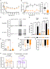
(A) von Frey thresholds for punctate allodynia were assessed for ipsilateral hindpaws in mice that received a single injection of anti-CD20 mAb or IgG2a isotype control (200 μg, intravenous), followed by CCI or sham surgery. Two-way repeated-measures ANOVA and Dunnett’s post hoc test; *P < 0.05, **P < 0.01, and ***P < 0.001 compared with sham/control. n = 11 to 12 per group (five or six males and six females). (B) Dynamic scores for ipsilateral hindpaws were determined within the same group of mice as in (A), using the brush test. The area under the curve (AUC) (arbitrary units, a.u.) was analyzed by Kruskal-Wallis and Dunn’s post hoc test; **P < 0.01 and ***P < 0.001. (C) Representative flow cytometry plots to confirm depletion of CD19+ B cells in bone marrow (BM), inguinal lymph node (iLN), popliteal lymph node (pLN), and spleen on days 3 and 49 after anti-CD20 mAb injection. (D) Proportion of CD19+ B cells at 3 and 49 days after injection. Two-way ANOVA and Šidák’s post hoc test; ***P < 0.001. n = 3 to 6 per group (one to three males and two or three females). (E) von Frey thresholds were assessed for the ipsilateral hindpaws in B cell–deficient muMT or wild-type (WT) mice that underwent CCI or sham surgery. Two-way repeated-measures ANOVA and Dunnett’s post hoc test; **P < 0.01 and ***P < 0.001 compared with sham/WT. n = 6 to 12 per group (sham groups = three males and three females; CCI groups = six males and six females). (F) Dynamic scores for the ipsilateral hindpaws were determined within the same group of mice as in (E). The AUC was analyzed by Kruskal-Wallis and Dunn’s post hoc test; **P < 0.01 and ***P < 0.001. (G) von Frey thresholds were assessed for ipsilateral hindpaws in muMT mice receiving adoptive transfer of 1.5 × 10⁷ B cells at 3 weeks before CCI. Baseline 1 (BL1) was measured before injections, and BL2 was measured 3 weeks after adoptive transfer. Two-way repeated-measures ANOVA and Šidák’s post hoc test; *P < 0.05. n = 6 per group (three males and three females). (H) Dynamic scores for ipsilateral hindpaws were determined within the same group of mice as in (G), using the brush test. The AUC was analyzed by Kruskal-Wallis and Dunn’s post hoc test; *P < 0.05.
Fig. 3.
Fig. 3.. IgG accumulation increases in mouse DRGs after peripheral nerve injury.
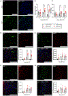
(A) Representative 40× fluorescent images of the ipsilateral lumbar DRGs 21 days after sham (top) or CCI (bottom) surgery. (B) Quantification of mean gray intensity for IgG staining in DRGs 3, 7, and 21 days after CCI or sham surgery. (C) Quantification of IgG protein concentration in DRGs by ELISA. Mean IgG concentrations from uninjured (naive) tissues are indicated by a dotted line. Two-way ANOVA and Tukey’s post hoc test; ***P < 0.001. n = 8 per group (four males and four females). (D to G) Representative 60× fluorescent images of ipsilateral lumbar DRGs 7 days after CCI surgery and quantification of colocalization of IgG with markers for (D) macrophages (F4/80), (E) neurons (MAP2), (F) satellite glia (GS), and (G) endothelial cells (CD31). Two-way ANOVA and Tukey’s post hoc test; *P < 0.05, **P < 0.01, and ***P < 0.001. n = 7 or 8 per group (three or four males and three or four females). Scale bars, 100 μm DAPI, 4′,6-diamidino-2-phenylindole.
Fig. 4.
Fig. 4.. IgG accumulation increases in the mouse spinal cord after peripheral nerve injury.
(A) Representative 40× fluorescent images of the ipsilateral lumbar dorsal horn 21 days after sham (top) or CCI (bottom) surgery. (B) Quantification of mean gray intensity for IgG staining in the lumbar dorsal horn 3, 7, and 21 days after CCI or sham surgery. (C) Quantification of IgG protein in the lumbar spinal cord by ELISA. Mean IgG concentrations from uninjured (naive) tissues are indicated by a dotted line. (D to G) Representative 60× fluorescent images of the ipsilateral lumbar dorsal horn 7 days after CCI surgery and quantification of colocalization of IgG with markers for (D) microglia (IBA1), (E) neurons (MAP2), (F) astrocytes (GFAP), and (G) endothelial cells (CD31). Two-way ANOVA and Tukey’s post hoc test; *P < 0.05, **P < 0.01, and ***P < 0.001. n = 7 or 8 per group (three or four males and three or four females). Scale bars, 100 μm.
Fig. 5.
Fig. 5.. IgG accumulation and B cell enrichment in DRGs from human donors with chronic pain.
(A) Representative 40× fluorescent images from human DRGs stained for IgG, nuclei (DAPI), markers for neurons [peripherin (PRPH)], and macrophages (GS). Scale bars, 100 μm. (B) Quantification of the fluorescence intensity for IgG in DRGs. Unpaired t test; **P < 0.01. n = 3 per group. See table S1 for donor demographics. (C) The extent of colocalization was quantified using the Manders overlap coefficient [proportion of pixels positive for IgG and markers for neurons (PRPH) or macrophages and satellite glia (GS) to pixels positive for IgG alone]. Two-way ANOVA and Tukey’s post hoc test; ***P < 0.001. (D) A transcriptional B cell signature in human DRGs derived by performing GSEA against a custom gene set for B cells from a recently published human cohort (34). The normalized enrichment score (NES; output from the ClusterProfiler package in R) was calculated per DRG using their ranked quantile-normalized transcripts per million counts. Shifts in the B cell NES were compared between patients with neuropathic pain (n = 33) and pain-free controls (n = 17) using a two-sample Kolmogorov-Smirnov test; *P < 0.05.
Fig. 6.
Fig. 6.. IgG from nerve-injured mice is pronociceptive.
(A) IgG from WT CCI or sham donor mice was passively transferred to CCI muMT recipient mice (three daily 40-μg intrathecal injections beginning 14 days after CCI). von Frey thresholds for punctate allodynia were assessed for ipsilateral hindpaws. Arrows indicate daily injections. Two-way ANOVA and Tukey’s post hoc test; *P < 0.05 and **P < 0.01. n = 12 males per group. (B) von Frey thresholds for punctate allodynia were assessed for ipsilateral hindpaws before (pre) and 24 hours after (post) the third daily injection of CCI IgG within the same group of mice as in (A). Paired t test. n = 18 males per group. (C) von Frey thresholds were assessed before (pre) and 24 hours after (post) the third daily injection of sham IgG within the same group of mice as in (A). Paired t test. n = 18 males per group. (D) IgG from WT CCI donor mice was passively transferred to naive muMT recipient mice (three daily 40-μg intrathecal injections). von Frey thresholds for punctate allodynia were assessed for left hindpaws before (pre) and 24 hours after (post) the third daily injection. Paired t test. n = 12 males per group.
Fig. 7.
Fig. 7.. FcγRs contribute to mechanical allodynia and DRG neuron hyperexcitability after peripheral nerve injury in mice.
(A) von Frey thresholds were assessed for the ipsilateral hindpaws of FcRγ−/− mice or WT littermates that had received CCI surgery. Two-way repeated-measures ANOVA and Šidák’s post hoc test; ***P < 0.001 compared with WT. n = 12 per group (six males and six females). (B) Dynamic scores for the ipsilateral hindpaws were determined within the same group of mice. The AUC was analyzed by the Mann-Whitney test; ***P < 0.001. (C) Representative traces from L4/5 DRG neurons dissociated from naive (Nv) WT mice (four males and two females), Nv FcRγ−/− (two males and two females), CCI WT (three males and three females), and CCI-FcRγ−/− mice (one male and three females) 14 days after CCI for patch clamp recording in current-clamp mode. Spontaneous activity (SA) was assessed at resting membrane potential (RMP), and ongoing activity was assessed when membrane potential was artificially held at −45 mV. (D) Incidence of DRG neurons with SA at RMP and ongoing activity when membrane potential was artificially held at −45 mV. (E) Rheobase (current threshold for initiating an action potential) with 2-s pulses, (F) RMP, and (G) action potential voltage thresholds. Data analyzed by two-sided Fisher’s exact test with Bonferroni correction for significance levels with multiple comparisons when comparing the incidence of neurons with ongoing activity and by one-way ANOVA and Dunnett’s post hoc test or Kruskal-Wallis and Dunn’s post hoc test; **P < 0.01 and ***P < 0.001. n = 12 to 15 neurons per group. (H) IgG from WT CCI donor mice was passively transferred to CCI-FcRγ−/− recipient mice (three daily 40-μg intrathecal injections beginning 14 days after CCI). von Frey thresholds for punctate allodynia were assessed for ipsilateral hindpaws before (pre) and 24 hours after (post) the third daily injection. Paired t test. n = 12 males per group. (I) F(ab′)2 fragments from CCI IgG were passively transferred to CCI muMT recipient mice (three daily 29-μg intrathecal injections). von Frey thresholds were assessed for ipsilateral hindpaws before (pre) and 24 hours after (post) the third daily injection. Paired t test. n = 9 males per group.

References

    1. van Hecke O, Austin SK, Khan RA, Smith BH, Torrance N, Neuropathic pain in the general population: A systematic review of epidemiological studies. Pain 155, 654–662 (2014). - PubMed
    1. Colloca L, Ludman T, Bouhassira D, Baron R, Dickenson AH, Yarnitsky D, Freeman R, Truini A, Attal N, Finnerup NB, Eccleston C, Kalso E, Bennett DL, Dworkin RH, Raja SN, Neuropathic pain. Nat. Rev. Dis. Primers 3, 17002 (2017). - PMC - PubMed
    1. Finnerup NB, Kuner R, Jensen TS, Neuropathic pain: From mechanisms to treatment. Physiol. Rev 101, 259–301 (2021). - PubMed
    1. Grace PM, Hutchinson MR, Maier SF, Watkins LR, Pathological pain and the neuroimmune interface. Nat. Rev. Immunol 14, 217–231 (2014). - PMC - PubMed
    1. McMahon SB, La Russa F, Bennett DL, Crosstalk between the nociceptive and immune systems in host defence and disease. Nat. Rev. Neurosci 16, 389–402 (2015). - PubMed

Publication types

LinkOut - more resources