. 2024 Oct 17;84(20):3932-3949.e10.
doi: 10.1016/j.molcel.2024.09.001. Epub 2024 Sep 24.

Structural basis of the human transcriptional Mediator regulated by its dissociable kinase module

Affiliations

Structural basis of the human transcriptional Mediator regulated by its dissociable kinase module

Ti-Chun Chao et al. Mol Cell. .

Abstract

The eukaryotic transcriptional Mediator comprises a large core (cMED) and a dissociable CDK8 kinase module (CKM). cMED recruits RNA polymerase II (RNA Pol II) and promotes pre-initiation complex formation in a manner repressed by the CKM through mechanisms presently unknown. Herein, we report cryoelectron microscopy structures of the complete human Mediator and its CKM. The CKM binds to multiple regions on cMED through both MED12 and MED13, including a large intrinsically disordered region (IDR) in the latter. MED12 and MED13 together anchor the CKM to the cMED hook, positioning CDK8 downstream and proximal to the transcription start site. Notably, the MED13 IDR obstructs the recruitment of RNA Pol II/MED26 onto cMED by direct occlusion of their respective binding sites, leading to functional repression of cMED-dependent transcription. Combined with biochemical and functional analyses, these structures provide a conserved mechanistic framework to explain the basis for CKM-mediated repression of cMED function.

Keywords: CDK8; CKM; CTD; IDR; MED12; MED13; MED26; PIC; RNA polymerase II; mediator; transcription.


Conflict of interest statement

Declaration of interests The authors declare no competing interests.

Figures
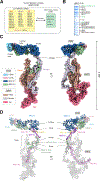
Figure 1.
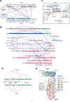
Figure 1.. Structure of the complete human Mediator
(A) Subunit composition of cMED and CKM, with the Metazoan-specific subunit MED26 in parentheses. (B) SDS-PAGE of purified Mediator. (C) Composite cryo-EM map of human cMED-CKM. The average resolutions for cMED and CKM are 4.7 Å and 6.7 Å, respectively. (D) Atomic model of human cMED-CKM with MED13 IDR bound to the Hinge of the Middle module. Two additional MED13 IDR binding sites on cMED are marked by green shapes.
Figure 2.
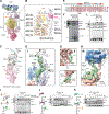
Figure 2.. Structure of the human CKM and its interaction with the cMED Hook
(A) Domain diagram of CKM subunits. (B) Composite cryo-EM map of human CKM. The resolutions of the Kinase-, Central-, and H-lobes are 3.8, 3.6, and 6.5 Å, respectively. (C) Structure of MED13 (colored) within the CKM. The missing MED13 IDR (residues 350–1069) is indicated by a dashed line. Other CKM subunits are colored in gray. (D) Two zinc-binding sites (I and II). Left, Zn2+ ion coordinated in MED13. Right, Zn2+ ion coordinated between MED13 and MED12. (E) Structure of MED12 (colored). (F) CKM bound to the cMED Hook. Bottom right: 2D average of CKM bound to Hook. (G) cMED Hook-CKM interface. (H) Conformational changes in the Kinase- and Central-lobes upon cMED binding. cMED-bound and free CKM structures were superimposed by aligning their H-lobes.
Figure 3.
Figure 3.. XL-MS analyses of Mediator and its CKM
(A) Crosslinking map and predicted IDRs within the human CKM. Intrasubunit and intersubunit URPs are shown by purple and blue lines, respectively. IDRs identified using IUPred3 are based on propensity scores, with residues above 0.5 considered disordered. Regions missing in density maps are colored white. (B) Crosslinking map of the cMED-CKM. (C) Crosslinks between MED12 H-lobe/MED13 and cMED Hook, with Cα-atoms shown as spheres. (D) Intrasubunit (purple) crosslinks within MED13/13L and their intersubunit crosslinks (green) with cMED subunits. (E) Crosslinks mapped between cMED and MED13/13L-IDR are projected onto the cMED-CKM structure.
Figure 4.
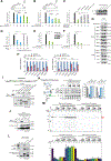
Figure 4.. MED13 uses its IDRc to interact with cMED
(A, C, E, G, and H) Pull-down analyses. (A) Flag-tagged MED13 immobilized on anti-Flag beads was used to pull-down cMED proteins from 293-F cell nuclear extract (NE). (B) MED13 fragments used for cMED interaction analysis. (C) Left, the N-terminal portion (residues 1–1320) of MED13 associates with cMED. Right, the IDRc, MBR1, and MBR2 of MED13 interact with cMED. (D) Glycerol gradient centrifugation analysis of purified MED13 IDRc-bound cMED. Gradient fractions were analyzed by SDS-PAGE with silver staining (top) and immunoblotting using indicated antibodies (bottom). (E) MED13/13L IDRc associate with cMED proteins from 293-F NE. (F) Mass spectrometric analysis. Heatmap showing scaled abundance of cMED, CKM, and Pol II proteins purified via Flag-tagged MED13 IDRc, CDK8, or MED26. (G) MED13 IDRc interacts with cMED, but not CKM subunits. MED13 with IDRc truncation cannot interact with cMED. (H) MED13 IDRc-bound cMED does not contain Pol II/MED26. GST-MED13 IDRc was used to capture cMED proteins isolated from HEK293 NE.
Figure 5.
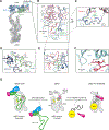
Figure 5.. MED13 IDRc excludes Pol II/MED26 interactions with cMED by occupying their binding sites
(A) The CKM main body does not overlap with Pol II/MED26 binding sites on cMED, but URPs (green) with MED13/13L IDRc do. (B) Crosslinks between cMED and MED13/13L IDRc are mapped onto the cMED-CKM structure. (C) Structure of the MED13 IDRc-bound cMED complex. Three IDRc binding sites (I, II, III) on cMED are indicated. (D) MED13 IDRc (green density) bound to the MED21/7 Hinge (I) of cMED. (E) Sequence alignment of a conserved region within the MBR2 of MED13 IDRc. (F) Pull-down experiment showing that GST-IDRc WT and Mut1, but not Mut2, interact with cMED proteins from 293-F NE. Mut1: F847D/S848Y/P849D. Mut2: M916D/F917D/A918Y. (G) MED13 IDRc bound to the Neck of Head module (II). (H) MED13 IDRc bridges the MED6 C-terminus and the Knob domain (III). (I-L) Competition binding assays. Protein concentrations (μM) used in competition are indicated. Eluted proteins were detected by western blotting using specified antibodies. (I-J) MED13 IDRc competes with Pol II-CTD or MED26 for cMED binding. (K) GST-CTD competes with MED13 IDRc for cMED binding. (L) WT MED13 IDRc, but not Mut2, competes with GST-CTD for cMED binding.
Figure 6.
Figure 6.. MED13 IDRc represses cMED-dependent transcription
(A-F) Luciferase reporter assays. Data plots represent relative luciferase activity from the average of triplicate independent transfections (*p < 0.05; **p < 0.01; ***p < 0.001). (A) Inhibition of Gal4-VP16-driven 5xGal4-TATA-luc (Gal4-Luc) reporter by MED13 and its IDRc. (B) Inhibition of Gal4-MED26-driven Gal4-Luc reporter by MED13 and its IDRc. (C) Inhibition of Gal4-MED26-driven Gal4-Luc reporter by WT MED13 IDRc and its mutants. (D) Inhibition of Gal4-MED26-driven Gal4-Luc reporter by WT MED13 and its (M916D/F917D/A918Y) mutant. (E and F) Inhibition of Gal4-MED26- or Gal4-VP16-driven Gal4-Luc reporter by MED13L IDRc. (G) Immunoblot analysis comparing expression levels of cMED and CKM subunits in the absence and presence of overexpressed MED13 IDRc in U2OS cells. (H) RT-qPCR analysis of genes regulated by the indicated transcription factors in human U2OS or ES cells transduced with control or MED13 IDRc-expressing lentiviruses. Relative expression levels are plotted based on three independent experiments. (I) Top, domain diagram of yeast Med13. Bottom, GST pull-down analysis using yeast Med13 fragments identifies one region (residues 563–700) within the IDR that binds cMed from whole cell extract (WCE). (J) GST pull-down analysis reveals interaction of WT, but not mutant, Med13 IDR with cMed from yeast WCE. yMut1: V609D/F610D/A611K; yMut2: G630R/G631R/K632E. (K) Competition binding assay. Yeast WT Med13 IDR, but not its mutants, competes with Pol II-CTD for cMed binding. Eluted proteins were detected by immunoblot, quantified, and normalized based on band intensities. (L) GST-tagged VP16 or Med13 IDR was used to pull-down cMed proteins from WCE. (M) Top, yeast in vitro transcription assay showing activator-dependent transcription repressed by CKM and WT Med13 IDR, but not by Med13 IDR mutants. Bottom, transcription products (indicated by a red arrow) were quantitated and normalized based on band intensities.
Figure 7.
Figure 7.. MED12-dependent CDK8 activation mechanism and proposed model for CKM-mediated transcription repression
(A) Position of CDK8/CCNC in the Mediator complex. (B) CDK8/CCNC bound by MED12N (blue), with the CDK8 T-loop in red and RHYT in green. (C) Human MED12N adopts a different conformation from yeast [PDB 7KPX] for CDK8 activation. Human MED12N is blue, yeast is white. (D) The RHYT segment (white) undergoes structural rearrangement (green) upon MED12N binding. (E) CDK8 T-loop and RHYT segment contacted by MED12N. (F) T-loop in the “DMG-Asp-in” conformation indicates active CDK8. (G) Model of CKM-mediated transcription repression. Before transcription initiation, CKM dispatches MED13 IDRc to block Pol II CTD/MED26 binding sites on cMED, inhibiting cMED-PIC formation and keeping cMED repressed. CKM dissociation permits Pol II/MED26 binding, enabling cMED-PIC formation and transcriptional activation. Tail module omitted for clarity.

Update of

References

    1. Kornberg RD (2005). Mediator and the mechanism of transcriptional activation. Trends Biochem Sci 30, 235–239. 10.1016/j.tibs.2005.03.011: . - DOI - PubMed
    1. Malik S, and Roeder RG (2010). The metazoan Mediator co-activator complex as an integrative hub for transcriptional regulation. Nat Rev Genet 11, 761–772. 10.1038/nrg2901: . - DOI - PMC - PubMed
    1. Soutourina J (2018). Transcription regulation by the Mediator complex. Nat Rev Mol Cell Biol 19, 262–274. 10.1038/nrm.2017.115: . - DOI - PubMed
    1. El Khattabi L, Zhao H, Kalchschmidt J, Young N, Jung S, Van Blerkom P, Kieffer-Kwon P, Kieffer-Kwon KR, Park S, Wang X, et al. (2019). A Pliable Mediator Acts as a Functional Rather Than an Architectural Bridge between Promoters and Enhancers. Cell 178, 1145–1158 e1120. 10.1016/j.cell.2019.07.011: . - DOI - PMC - PubMed
    1. Adelman K, and Lis JT (2012). Promoter-proximal pausing of RNA polymerase II: emerging roles in metazoans. Nat Rev Genet 13, 720–731. 10.1038/nrg3293: . - DOI - PMC - PubMed

MeSH terms

LinkOut - more resources