. 2024 Oct;300(10):107813.
doi: 10.1016/j.jbc.2024.107813. Epub 2024 Sep 23.

Stub1 promotes degradation of the activated Diaph3: A negative feedback regulatory mechanism of the actin nucleator

Affiliations

Stub1 promotes degradation of the activated Diaph3: A negative feedback regulatory mechanism of the actin nucleator

Cui Qiu et al. J Biol Chem. 2024 Oct.

Abstract

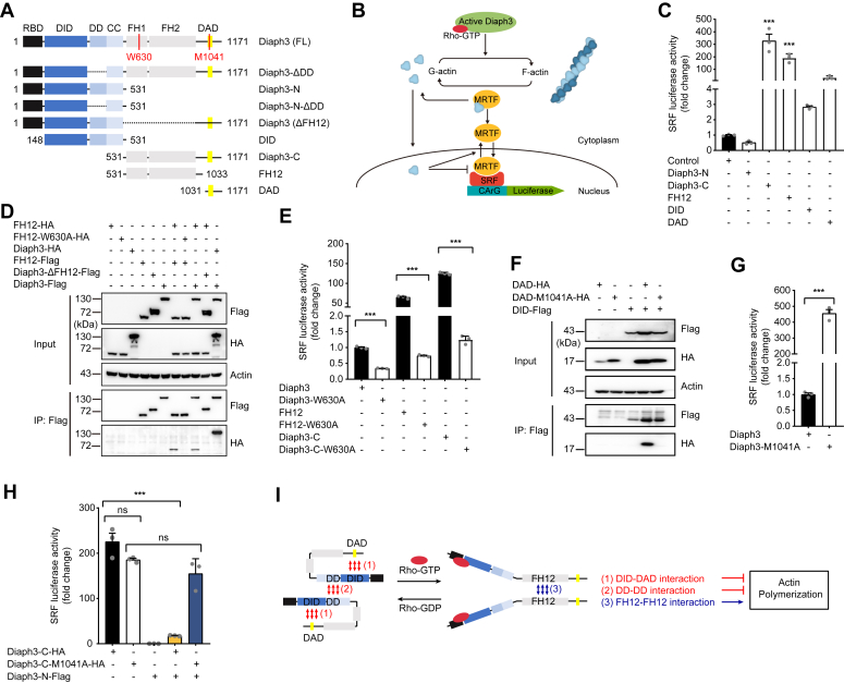
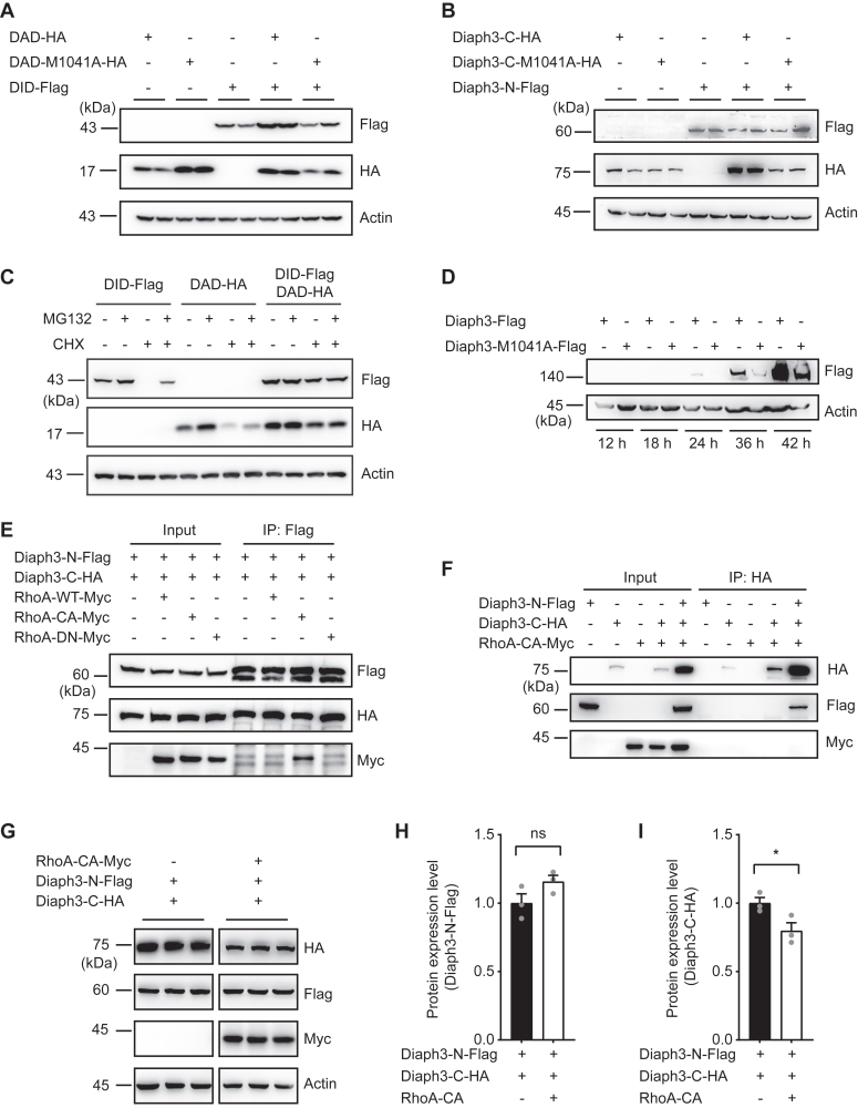
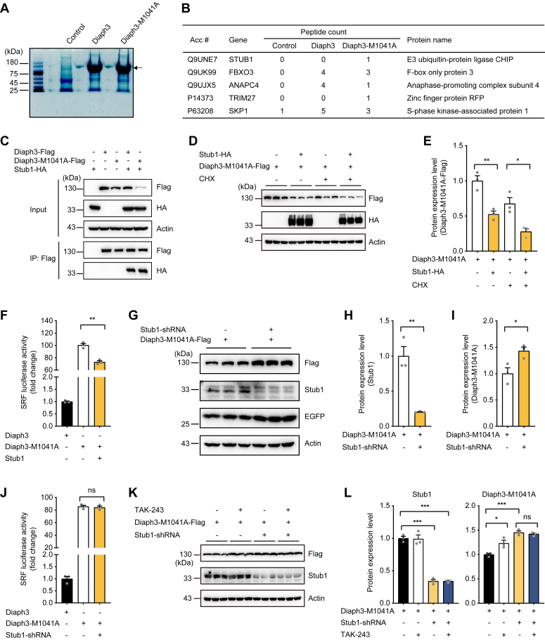
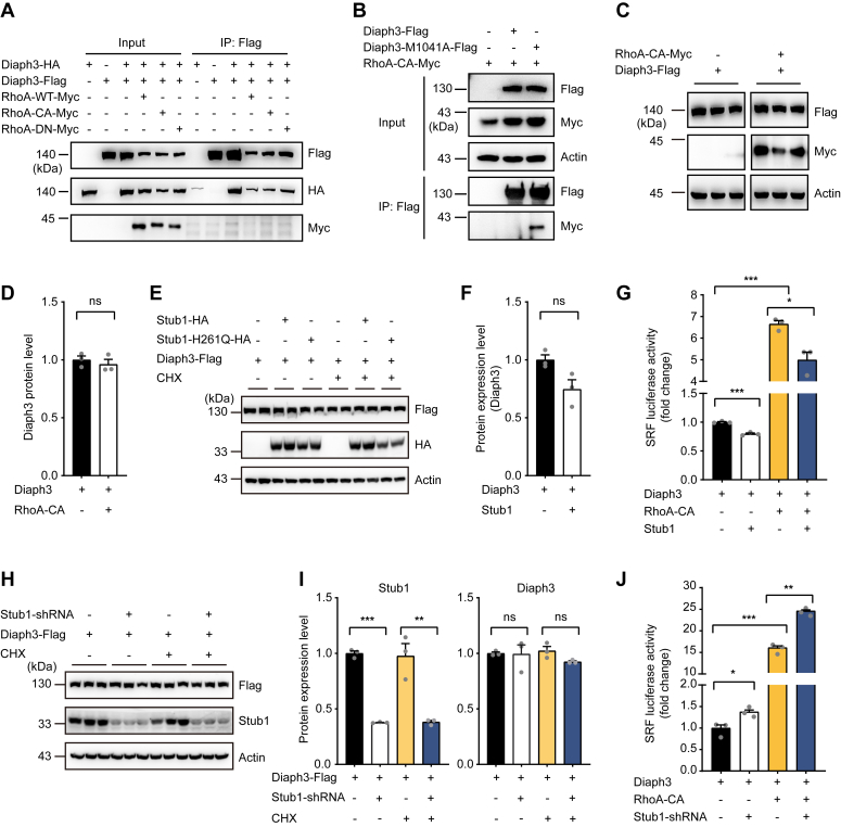
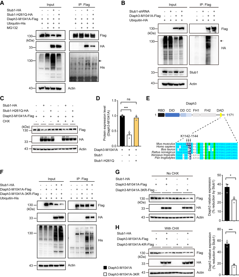
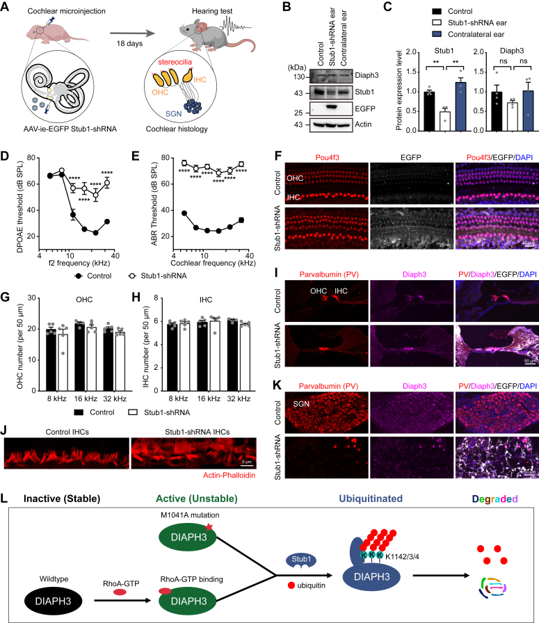
The formin protein Diaph3 is an actin nucleator that regulates numerous cytoskeleton-dependent cellular processes through the activation of actin polymerization. Expression and activity of Diaph3 is tightly regulated: lack of Diaph3 results in developmental defects and embryonic lethality in mice, while overexpression of Diaph3 causes auditory neuropathy. It is known that Diaph3 homophilic interactions include the intramolecular interaction of its Dia-inhibitory domain (DID)-diaphanous autoregulatory domain (DAD) domains and the intermolecular interactions of DD-DD domains or FH2-FH2 domains. However, the physiological significance of these interactions in Diaph3 protein stability and activity is not fully understood. In this study, we show that FH2-FH2 interaction promotes Diaph3 activity, while DID-DAD and DD-DD interactions inhibit Diaph3 activity through distinct mechanisms. DID-DAD interaction is responsible for the autoinhibition of Diaph3 protein, which is disrupted by binding of Rho GTPases. Interestingly, we find that DID-DAD interaction stabilizes the expression of each DID or DAD domain against proteasomal-mediated degradation. Disruption of DID-DAD interaction by RhoA binding or M1041A mutation causes increased Diaph3 activity and accelerated degradation of the activated Diaph3 protein. Further, the activated Diaph3 is ubiquitinated at K1142/1143/1144 lysine residues by the E3 ligase Stub1. Expression of Stub1 is causally related to the stability and activity of Diaph3. Knockdown of Stub1 in mouse cochlea results in hair cell stereocilia defects, neuronal degeneration, and hearing loss, resembling the phenotypes of mice overexpressing Diaph3. Thus, our study reports a novel regulatory mechanism of Diaph3 protein expression and activity whereby the active but not inactive Diaph3 is readily degraded to prevent excessive actin polymerization.

Keywords: Diaph3; Stub1; actin polymerization; autoinhibition; hearing loss; negative feedback; protein stability.


Conflict of interest statement

Conflict of interest The authors declare that they have no conflicts of interest with the contents of this article.

Figures

Figure 1
Figure 1
Diaph3 activity is regulated by its intramolecular and intermolecular interactions.A, schematic diagram of Diaph3 domains and constructs. B, schematic diagram of actin polymerization activity assayed by the SRF-RE luciferase reporter system. C, SRF activity of the truncated Diaph3 constructs transfected in HEK293T cells. D, intermolecular interaction of the FH12-FH12 domains was inhibited by the W630A mutation. E, intermolecular interaction of the FH12-FH12 domains was required for Diaph3 activity. F, intramolecular DID-DAD domain interaction (autoinhibition) was disrupted by the M1041A mutation. G, mutation of M1041A in full-length Diaph3 significantly increased SRF activity. H, interaction of Diaph3-C and Diaph3-N inhibited the SRF activity of Diaph3-C. I, actin polymerization activity of Diaph3 protein induced by binding to the active Rho GTPase is mediated by intermolecular FH12 interactions, but inhibited by both intramolecular DID-DAD interactions and intermolecular DD-DD interactions. Mean ± SEM. ns, not significant, ∗∗∗p < 0.001 by unpaired Student’s t test. DAD, diaphanous autoregulatory domain; DD, dimerization domain; DID, Dia-inhibitory domain; SRF, serum response factor.
Figure 2
Figure 2
DID-DAD interaction-mediated autoinhibition promotes Diaph3 protein stabilization.A, protein stability of DID and DAD constructs was increased by DID-DAD interaction, and was abolished by M1041A mutation. Western blots of HEK293T cells with DAD-HA, DAD-M1041A-HA, or DID-Flag for Flag, HA and actin. B, Diaph3-N stabilized the expression of Diaph3-C but not Diaph3-C-M1041A. C, autoinhibition between DID and DAD increased protein stability via proteasomal degradation pathways. Cells were treated with 10 μM MG132 (proteasomal inhibitor) and/or 10 μM cycloheximide (CHX) for 4 h. D, M1041A mutation promoted degradation of the full-length Diaph3. E, RhoA-CA interacted with N-terminal fragment of Diaph3. WT, wildtype; CA, constitutively active; DN, dominant negative. F, C-terminal fragment of Diaph3 did not interact with RhoA-CA. Western blots of anti-HA immunoprecipitation of HA-Flag-Myc and whole cell lysate for Flag, HA, Myc from HEK293T cells. G, RhoA-CA reduced the stabilization of Diaph3 C-terminal fragment. H and I, protein quantification of Diaph3-N-Flag and Diaph3-C-HA in panel G. Mean ± SD. ns, not significant, ∗p < 0.05 by unpaired Student’s t test. DAD, diaphanous autoregulatory domain; DID, Dia-inhibitory domain; HA, hemagglutinin.
Figure 3
Figure 3
E3 ubiquitin ligase Stub1 regulates the degradation of Diaph3-M1041A.A, coomassie blue gel staining of MG132-treated immunoprecipitated Diaph3 proteins for mass spectrometry. The arrow indicates the Diaph3 protein (140 kD). B, list of Diaph3-interacting E3 ubiquitin ligases from mass spectrometry data. C, WT Diaph3 and Diaph3-M1041A interacted with the E3 ubiquitin ligase Stub1. D and E, Western blots (D) and quantitative data (E) showed that Stub1 overexpression promoted the degradation of Diaph3-M1041A with or without CHX treatment. Mean ± SD, ∗p < 0.05, ∗∗p < 0.01 by unpaired Student’s t test. F, Stub1 overexpression decreased SRF activity of Diaph3-M1041A. Mean ± SEM. ∗∗p < 0.01 by unpaired Student’s t test. G, Stub1 knockdown increased Diaph3-M1041A expression. H and I, Protein quantification of Stub1 (H) and Diaph3-M1041A (I) in panel G. Mean ± SD, ∗p < 0.05, ∗∗p < 0.01 by unpaired Student’s t test. J, Stub1 knockdown did not affect SRF activity of Diaph3-M1041A. Mean ± SEM. ns, not significant. K, The E1 inhibitor TAK-243 did not further increase Diaph3-M1041A protein levels after Stub1 knockdown. Cells were treated with 5 μM TAK-243 for 6 h. L, Protein quantification of Stub1 and Diaph3-M1041A in panel K. Mean ± SD. ns, not significant, ∗p < 0.05, ∗∗∗p < 0.001 by unpaired Student’s t test. CHX, cycloheximide; SRF, serum response factor.
Figure 4
Figure 4
Stub1 modulates the activity of WT Diaph3.A, RhoA did not interact with full length Diaph3. B, Diaph3-M1041A but not full length Diaph3 interacted with RhoA-CA. C, RhoA did not affect protein stability of the full length Diaph3. D, protein quantification of Diaph3-Flag in panel C. Mean ± SD. ns, not significant. E, Stub1 overexpression did not affect the expression of WT Diaph3 with or without CHX treatment. F, protein quantification of Diaph3-Flag after Stub1 overexpression. Mean ± SD. ns, not significant. G, Stub1 overexpression decreased SRF activity of the WT Diaph3 in the presence or absence of RhoA-CA. Mean ± SEM. ∗p < 0.05, ∗∗∗p < 0.001 by unpaired Student’s t test. H, Stub1 knockdown did not affect the expression of WT Diaph3 with or without CHX treatment. I, protein quantification of Stub1 and Diaph3-Flag in panel H. Mean ± SD. ns, not significant, ∗∗p < 0.01, ∗∗∗p < 0.001 by unpaired Student’s t test. J, Stub1 knockdown increased SRF activity of the WT Diaph3 in the presence or absence of RhoA-CA. Mean ± SEM. ∗p < 0.05, ∗∗p < 0.01, ∗∗∗p < 0.001 by unpaired Student’s t test. SRF, serum response factor.
Figure 5
Figure 5
Stub1 mediates polyubiquitination of Diaph3-M1041A.A, overexpression WT Stub1 but not Stub1-H261Q mutant induced Diaph3-M1041A polyubiquitination. The arrow indicates ubiquitin covalently bound to the lysine residues of Diaph3-M1041A (140 kD). B, Stub1 knockdown reduced Diaph3-M1041A polyubiquitination. C, overexpression of WT Stub1 but not Stub1-H261Q mutant reduced Diaph3-M1041A protein expression with or without CHX treatment. D, protein quantification of Diaph3-M1041A-Flag after Stub1 or Stub1-H261Q overexpression. E, conservation of the Diaph3 K1142 to 1144 residues in different mammalian species. F, the 3 KR mutation reduced Diaph3-M1041A polyubiquitination by Stub1. G and H, Western blots and quantitative data showed that the 3KR mutation partially counteracted the decrease in Diaph3-M1041A expression caused by Stub1 overexpression without CHX (G) or with CHX (H). Mean ± SD. ns, not significant, ∗p < 0.05, ∗∗∗p < 0.001 by unpaired Student’s t test.
Figure 6
Figure 6
Knockdown of the endogenous Stub1 in mouse inner ear leads to severe hearing loss.A, schematic diagram of knocking down endogenous Stub1 expression in mouse inner ear. AAV-ie vector containing GFP tag was used to package Stub1-shRNA. The left ear of P3 mice were injected with AAV-ie via round window membrane (RWM). Hearing measurements was performed 18 days (P21) after injection. Cochlear histology was performed to examine survival and morphology of hair cells, stereocilia bundles and spiral ganglion neurons (SGNs). IHC, inner hair cell; OHC, outer hair cell. B and C, Western blot images (B) and protein quantification (C) of Stub1 and Diaph3 expression in control and Stub1 knockdown cochlea. Mean ± SD. ns, not significant, ∗∗p < 0.01 by unpaired Student’s t test. D and E, DPOAE (D) and ABR (E) thresholds in control and Stub1 knockdown mice at P21. n = 18 ears each condition. Mean ± SEM. ∗∗∗∗p < 0.0001 by two-way ANOVA followed by Bonferroni's post test. SPL, sound pressure level. ABR, auditory brainstem response; DPOAE, distortion product otoacoustic emission. F, wholemount immunofluorescent images of hair cells labeled with anti-Pou4f3 antibody (red) in control and Stub1 knockdown cochlea at P21. G and H, numbers of outer hair cells (G) and inner hair cells (H) at different cochlear frequencies (8, 16, 32 kHz) (n = 5 cochleae each condition). Mean ± SEM. I, immunofluorescent images showing Parvalbumin (hair cells) and Diaph3 expression in cryo-sectioned cochlea (organ of Corti area). J, hair cell stereocilia bundle morphology labeled with Actin-Phalloidin. K, immunofluorescent images showing Parvalbumin (SGNs) and Diaph3 expressions in cryo-sectioned cochlea (SGN area). L, proposed mechanism of a novel negative feedback regulation of Diaph3 protein expression and activity by Stub1-mediated degradation. AAV-ie, AAV-inner ear.

Similar articles

References

    1. Evangelista M., Pruyne D., Amberg D.C., Boone C., Bretscher A. Formins direct Arp2/3-independent actin filament assembly to polarize cell growth in yeast. Nat. Cell Biol. 2002;4:260–269. - PubMed
    1. Feierbach B., Chang F. Roles of the fission yeast formin for3p in cell polarity, actin cable formation and symmetric cell division. Curr. Biol. 2001;11:1656–1665. - PubMed
    1. Kobielak A., Pasolli H.A., Fuchs E. Mammalian formin-1 participates in adherens junctions and polymerization of linear actin cables. Nat. Cell Biol. 2004;6:21–30. - PMC - PubMed
    1. Severson A.F., Baillie D.L., Bowerman B. A Formin Homology protein and a profilin are required for cytokinesis and Arp2/3-independent assembly of cortical microfilaments in C. elegans. Curr. Biol. 2002;12:2066–2075. - PubMed
    1. Miralles F., Posern G., Zaromytidou A.I., Treisman R. Actin dynamics control SRF activity by regulation of its coactivator MAL. Cell. 2003;113:329–342. - PubMed

Publication types

LinkOut - more resources