. 2024 Oct 15:300:120863.
doi: 10.1016/j.neuroimage.2024.120863. Epub 2024 Sep 23.

Dynamic functional connectivity in verbal cognitive control and word reading

Affiliations

Dynamic functional connectivity in verbal cognitive control and word reading

Kazuki Sakakura et al. Neuroimage. .

Abstract

Cognitive control processes enable the suppression of automatic behaviors and the initiation of appropriate responses. The Stroop color naming task serves as a benchmark paradigm for understanding the neurobiological model of verbal cognitive control. Previous research indicates a predominant engagement of the prefrontal and premotor cortex during the Stroop task compared to reading. We aim to further this understanding by creating a dynamic atlas of task-preferential modulations of functional connectivity through white matter. Patients undertook word-reading and Stroop tasks during intracranial EEG recording. We quantified task-related high-gamma amplitude modulations at 547 nonepileptic electrode sites, and a mixed model analysis identified regions and timeframes where these amplitudes differed between tasks. We then visualized white matter pathways with task-preferential functional connectivity enhancements at given moments. Word reading, compared to the Stroop task, exhibited enhanced functional connectivity in inter- and intra-hemispheric white matter pathways from the left occipital-temporal region 350-600 ms before response, including the posterior callosal fibers as well as the left vertical occipital, inferior longitudinal, inferior fronto-occipital, and arcuate fasciculi. The Stroop task showed enhanced functional connectivity in the pathways from the left middle-frontal pre-central gyri, involving the left frontal u-fibers and anterior callosal fibers. Automatic word reading largely utilizes the left occipital-temporal cortices and associated white matter tracts. Verbal cognitive control predominantly involves the left middle frontal and precentral gyri and its connected pathways. Our dynamic tractography atlases may serve as a novel resource providing insights into the unique neural dynamics and pathways of automatic reading and verbal cognitive control.

Keywords: Dynamic tractography; Executive function; Functional brain mapping; Language; Pediatric epilepsy surgery; Physiological high-frequency oscillation (HFO).


Conflict of interest statement

Declaration of competing interest None.

Figures

Fig. 1.
Fig. 1.
Spatial extent of non-epileptic electrode sites on a standard brain surface image. (A) This figure indicates the number of patients available at a given analysis mesh. (B) This figure indicates the locations of region of interests (ROIs) on the left hemisphere. aCG: anterior cingulate gyrus. aFG: anterior fusiform gyrus. aIFG: anterior inferior-frontal gyrus. aITG: anterior inferior-temporal gyrus. aMFG: anterior middle-frontal gyrus. Cun: cuneus. Ent: entorhinal gyrus. FP: frontal pole. IPL: inferior parietal lobule. LG: lingual gyrus. LOG: lateral occipital gyrus. MOrb: medial orbitofrontal gyrus. MTG: middle-temporal gyrus. pCG: posterior cingulate gyrus. PCl: pericalcarine cortex. PCL: paracentral lobule. PCun: precuneus. pFG: posterior fusiform gyrus. PHG: parahippocampal gyrus. pIFG: posterior inferior-frontal gyrus. pITG: posterior inferior-temporal gyrus. pMFG: posterior middle-frontal gyrus. PoCG: postcentral gyrus. PreCG: precentral gyrus. SFG: superior-frontal gyrus. SMG: supramarginal gyrus. SPL: superior parietal lobule. STG: superior-temporal gyrus. TP: temporal pole. Group-level regions of interest (ROIs) were employed only in those with at least five nonepileptic electrode sites. The number of non-epileptic electrode sites in each ROI is provided in Table S1.
Fig. 2.
Fig. 2.
Tasks. (A) Congruent word reading task. (B) Incongruent word reading task. (C) Stroop color naming task. The response time is characterized as the period between the onset of the stimulus and the onset of the response. The baseline period for intracranial EEG analysis pertains to the 400-ms quiet period between 1800 and 2200 ms post-response onset.
Fig. 3.
Fig. 3.
Task-related high-gamma amplitude modulations. The snapshots showcase the percent change of high-gamma amplitudes in comparison to the baseline mean. Upper: high-gamma modulations during word reading (i.e., average during congruent and incongruent word-reading tasks). Arrows indicate high-gamma augmentation in the posterior fusiform region. Lower: Stroop color naming. An arrowhead indicates high-gamma augmentation in the posterior middle frontal gyrus. For a comprehensive overview of the high-gamma amplitude changes during each task, please refer to Video S1.
Fig. 4.
Fig. 4.
Task-related modulations of high-gamma amplitude. A given plot presents the percent change in high-gamma amplitudes at each anatomical ROI compared to the baseline mean at 1800 to 2200 ms after response onset. Magenta: word-reading (i.e., average of congruent and incongruent word reading tasks). Green: Stroop color-naming. Shade: 95 % confidence interval.
Fig. 5.
Fig. 5.
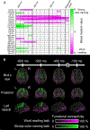
Cortical and white matter substrates preferentially supporting given tasks. (A) Spatiotemporal characteristics of high-gamma amplitudes differentially modulated during Stroop color naming versus word reading. Cells present mixed model t-values for specific regions of interest (ROIs) and their corresponding 200-ms time windows (e.g., −600 ms: a time window between −600 and −400 ms pre-response onset). These colors indicate when and where high-gamma amplitude was significantly higher (green) or lower (magenta) during Stroop color naming relative to word reading. Readers will find that high-gamma amplitude at the left posterior middle-frontal gyrus (pMFG) was higher during Stroop task compared to word reading at two time windows starting at −600 ms and −550 ms pre-response (i.e., between −600 and −400 ms and −550 and −350 ms before verbal responses). (B) Color-coded streamlines depict the spatiotemporal dynamics of functional connectivity enhancement between ROIs preferential to Stroop color naming (green) and word reading (magenta). For a comprehensive overview of the task-preferential network dynamics, please refer to Video S2. aIFG: anterior inferior-frontal gyrus. aITG: anterior inferior-temporal gyrus. aMFG: anterior middle-frontal gyrus. Cun: cuneus. IPL: inferior parietal lobule. LG: lingual gyrus. LOG: lateral occipital gyrus. MTG: middle-temporal gyrus. PCun: precuneus. pFG: posterior fusiform gyrus. pIFG: posterior inferior-frontal gyrus. pITG: posterior inferior-temporal gyrus. PoCG: postcentral gyrus. PreCG: precentral gyrus. SFG: superior-frontal gyrus. SMG: supramarginal gyrus. STG: superior-temporal gyrus. TP: temporal pole.

References

    1. Adleman NE, Menon V, Blasey CM, White CD, Warsofsky IS, Glover GH, Reiss AL, 2002. A developmental fMRI study of the Stroop color-word task. Neuroimage 16, 61–75. 10.1006/nimg.2001.1046. - DOI - PubMed
    1. Alexander MP, Stuss DT, Picton T, Shallice T, Gillingham S, 2007. Regional frontal injuries cause distinct impairments in cognitive control. Neurology 68, 1515–1523. 10.1212/01.wnl.0000261482.99569.fb. - DOI - PubMed
    1. Asano E, Juhász C, Shah A, Sood S, Chugani HT, 2009. Role of subdural electrocorticography in prediction of long-term seizure outcome in epilepsy surgery. Brain 132, 1038–1047. 10.1093/brain/awp025. - DOI - PMC - PubMed
    1. Asato MR, Terwilliger R, Woo J, Luna B, 2010. White matter development in adolescence: a DTI study. Cereb. Cortex 20, 2122–2131. 10.1093/cercor/bhp282. - DOI - PMC - PubMed
    1. Ball T, Demandt E, Mutschler I, Neitzel E, Mehring C, Vogt K, Aertsen A, Schulze-Bonhage A, 2008. Movement related activity in the high gamma range of the human EEG. Neuroimage 41, 302–310. 10.1016/j.neuroimage.2008.02.032.. - DOI - PubMed

LinkOut - more resources