

Temporal progression of sleep electroencephalography features in isolated rapid eye movement sleep behaviour disorder
- PMID: 39322419
- PMCID: PMC12069751
- DOI: 10.1111/jsr.14351
Temporal progression of sleep electroencephalography features in isolated rapid eye movement sleep behaviour disorder
Abstract
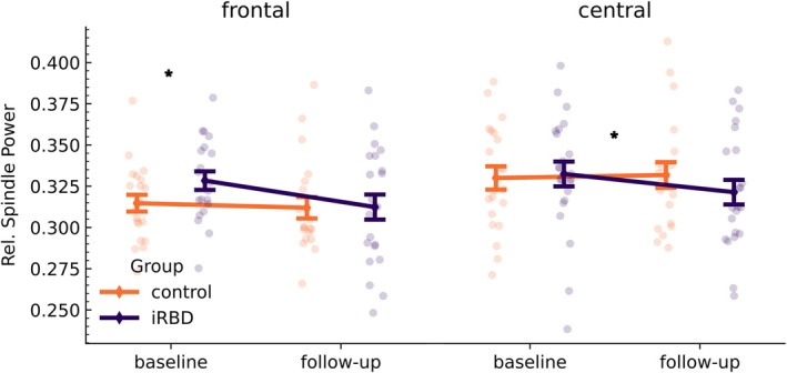
Previous studies indicated that patients with isolated rapid eye movement (REM) sleep behaviour disorder (iRBD) exhibit alterations in spectral electroencephalographic (EEG), spindle, and slow-wave features. As it is currently unknown how these EEG features evolve over time, this study aimed to evaluate their temporal progression in patients with iRBD in comparison to controls. We included 23 patients with iRBD and 23 controls. Two polysomnographies (baseline and follow-up) were recorded with a mean (standard deviation) interval of 4.0 (2.5) years and were automatically analysed for sleep stages, spectral bandpower, spindles, and slow waves. We used linear models to evaluate differences at each time point, and linear mixed-effects models to analyse differences in temporal progression between the groups. At baseline, patients with iRBD presented EEG slowing both in REM (expressed as significantly reduced α-bandpower and increased δ-bandpower in frontal channels) and in non-REM (NREM) sleep (significantly increased slow-to-fast ratio in central channels). These differences vanished at follow-up. In both REM and NREM sleep, γ-bandpower was increased at follow-up in patients with iRBD, resulting in significantly different temporal progression between groups (in occipital channels during REM sleep and frontal channels during NREM sleep). Relative power of sleep spindles was significantly higher at baseline in patients with iRBD in frontal channels, but we observed a significant reduction over time in central channels. Finally, slow waves were significantly shorter in patients with iRBD at both time-points. Our results underscore the need of considering longitudinal data when analysing sleep EEG features in patients with iRBD. The observed temporal changes as markers of progression of neurodegeneration require further investigations.
Keywords: computerised methods; electrophysiology; prodromal alpha‐synucleinopathy.
© 2024 The Author(s). Journal of Sleep Research published by John Wiley & Sons Ltd on behalf of European Sleep Research Society.
Conflict of interest statement
None.
Figures


References
-
- Althouse, A. D. (2016). Adjust for multiple comparisons? It's not that simple. The Annals of Thoracic Surgery, 101(5), 1644–1645. - PubMed
-
- American Academy of Sleep Medicine (Ed.). (2014). The International Classification of Sleep Disorders (ICSD‐3) (3rd ed.). American Academy of Sleep Medicine.
-
- Bender, R. , & Lange, S. (2001). Adjusting for multiple testing—when and how? Journal of Clinical Epidemiology, 54(4), 343–349. - PubMed
-
- Berry, R. , Brooks, R. , Gamaldo, C. , Harding, S. , Marcus, C. , Vaughn, B. , & the American Academy of School Medicine . (2012). The AASM Manual for the Scoring of Sleep and Associated Events: Rules, Terminology and Technical Specifications. Version 2.0. 2nd ed. Darien, IL.
-
- Berry, R. , Brooks, R. , Gamaldo, C. , Harding, S. , Marcus, C. , Vaughn, B. , & the American Academy of Sleep Medicine . (2020). The AASM Manual for the Scoring of Sleep and Associated Events: Rules, Terminology and Technical Specifications. Version 2.6. 2nd ed. Darien, IL.
Publication types
MeSH terms
Grants and funding
LinkOut - more resources
Full Text Sources
Research Materials
