. 2025 Apr;42(2):398-409.
doi: 10.1007/s12028-024-02133-9. Epub 2024 Sep 25.

The Pupillary Light-Off Reflex in Acute Disorders of Consciousness

Affiliations

The Pupillary Light-Off Reflex in Acute Disorders of Consciousness

Pardis Zarifkar et al. Neurocrit Care. 2025 Apr.

Abstract

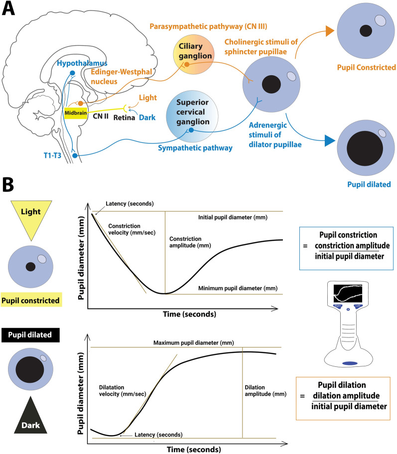
Background: In intensive care patients with disorders of consciousness, the pupillary light reflex is a measure of pupillary parasympathetic function. By contrast, the pupillary light-off reflex leads to pupil dilation in response to an abrupt change from light to darkness ("light-off") and reflects combined parasympathetic and sympathetic pupillary function. To our knowledge, this reflex has not been systematically investigated in patients with disorders of consciousness. We hypothesized that the pupillary light-off reflex correlates with consciousness levels after acute brain injury.

Methods: From November 2022 to March 2023, we enrolled 100 study participants: 25 clinically unresponsive (coma or unresponsive wakefulness syndrome) and 25 clinically low-responsive (minimally conscious state or better) patients from the intensive care units of a tertiary referral center, and 50 age-matched and sex-matched healthy controls. Exclusion criteria were active or chronic eye disease. We used automated pupillometry to assess the pupillary light-off reflex and the pupillary light reflex of both eyes under scotopic conditions in all study participants.

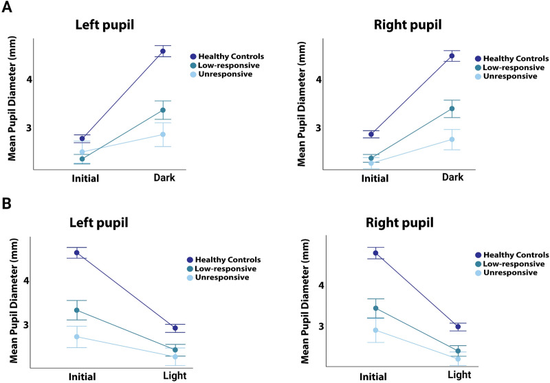
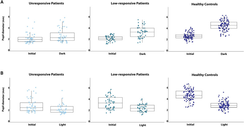
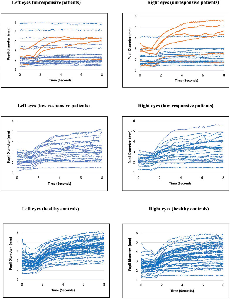
Results: The pupillary light-off reflex was strongly correlated with consciousness levels (r = 0.62, p < 0.001), the increase in pupillary diameters being smallest in unresponsive patients (mean ± standard deviation 20% ± 21%), followed by low-responsive patients (mean ± standard deviation 47% ± 26%) and healthy controls (mean ± standard deviation 67% ± 17%; p < 0.001). Similar yet less pronounced patterns were observed for the pupillary light reflex. Twenty-one of 25 (84%) unresponsive patients had preserved pupillary light reflexes, but only seven (28%) had fully preserved pupillary light-off reflexes (p < 0.0001). Of these 7 patients, five (71%) regained awareness.

Conclusions: The pupillary light-off reflex may be more sensitive to consciousness levels than the pupillary light reflex. The clinical implications of this finding seem worthy of further investigation, particularly regarding possible benefits for neuromonitoring and prognostication after brain injury.

Keywords: Brain injury; Coma; Consciousness; Neuromonitoring; Prognostication; Pupillometry.


Conflict of interest statement

Conflicts of interest: The authors report no competing interests. Ethical approval/informed consent: The study adhered to the Declaration of Helsinki and received approval from the regional Danish Research Ethics Committee (H-21022096). Ethical guidelines were adhered to, and we indicate ethical approvals (institutional review board) and use of informed consent, and we confirm the use of an appropriate reporting checklist.

Figures

Fig. 1
Fig. 1
Simplified illustration of some of the parasympathetic and sympathetic pathways that regulate a pupil size and b measurement of the pupillary light and light-off reflexes using automated pupillometry. When exposed to light, retinal cells send signals through the optic nerve to the pretectal nucleus, which communicates with the Edinger–Westphal nucleus. Parasympathetic fibers travel via the oculomotor nerve to the ciliary ganglion and the sphincter pupillae muscle, causing pupil constriction (pupillary light reflex). In darkness, the retina signals the hypothalamus, activating the sympathetic pathway via the spinal cord (T1-T2), leading to the superior cervical ganglion. Postganglionic fibers then travel to the dilator pupillae muscle, resulting in pupil dilation (pupillary light-off reflex). The pupillary light reflex is the difference between initial pupillary diameter and the minimal pupillary diameter after constriction, divided by the initial pupil diameter. By contrast, the pupillary light-off reflex is the difference between the initial pupillary diameter and the maximal pupillary diameter after dilatation, divided by the initial pupil diameter. A change in pupillary diameter during pupillary dilation or constriction is defined as the increase or decrease in pupillary diameter (curve amplitude) relative to the initial diameter. It should be noted that the figure omits the contribution of the inhibitory effect on the Edinger–Westphal nucleus and that the anatomical pathways mediating the pupillary light-off reflex are not yet entirely understood
Fig. 2
Fig. 2
Pupillary diameter changes as a function of a the pupillary light-off reflex and b the pupillary light reflex. The figures illustrate changes in mean pupillary diameter during the pupillary light-off and light reflexes for unresponsive patients, low-responsive patients, and healthy controls in left and right eyes. Patients exhibited significantly smaller increases and decreases in pupillary diameter compared to healthy controls, with unresponsive patients showing the smallest changes. Like the pupillary light reflex, the pupillary light-off reflects distinguishes well between clinically unresponsive and low-responsive patients. For individual pupillary diameter data see Fig. 3
Fig. 3
Fig. 3
Individual pupillary diameter changes during a the pupillary light-off reflex and b the pupillary light reflex for clinically unresponsive patients, low-responsive patients, and healthy controls, displayed as scatter plots with overlaying box plots
Fig. 4
Fig. 4
Raw data of the pupillary light-off reflex of clinically unresponsive and low-responsive patients, and age-and sex-matched healthy controls, illustrating pupillary diameters over time. Time in seconds is displayed on x-axes, and pupillary diameters in millimeters are shown on y-axes. Preserved light-off reflexes of unresponsive patients, as defined by pupillary dilation within two standard deviations of the mean pupillary dilation response observed in healthy controls, are highlighted in orange. Note that rarely, a rudimentary pupillary dilation response appears in unresponsive patients that is outside the normal range. The figure shows that the shape and distribution of the pupillary light-off reflex correlate with increasing consciousness levels, i.e., from mostly absent in clinically unresponsive patients to brisk in healthy volunteers

Similar articles

Cited by

References

    1. Peinkhofer C, Martens P, Grand J, et al. Influence of strategic cortical infarctions on pupillary function. Front Neurol. 2018;9:916. - PMC - PubMed
    1. Greer DM, Yang J, Scripko PD, et al. Clinical examination for prognostication in comatose cardiac arrest patients. Resuscitation. 2013;84:1546–51. - PMC - PubMed
    1. Loewenfeldt I. The pupil: anatomy, physiology, and clinical applications. 2nd ed. Oxford: Butterworth-Heinemann; 1999.
    1. Hunyor AP. Reflexes and the eye. Aust N Z J Ophthalmol. 1994;22:155–9. - PubMed
    1. Cherng Y-G, Crevecoeur F, Wang C-A. Effects of pupillary light and darkness reflex on the generation of pro- and anti-saccades. Eur J Neurosci. 2021;53:1769–82. - PubMed