. 2024 Dec;37(12):e5262.
doi: 10.1002/nbm.5262. Epub 2024 Sep 25.

DCE-MRI of the liver with sub-second temporal resolution using GRASP-Pro with navi-stack-of-stars sampling

Affiliations

DCE-MRI of the liver with sub-second temporal resolution using GRASP-Pro with navi-stack-of-stars sampling

Jingjia Chen et al. NMR Biomed. 2024 Dec.

Abstract

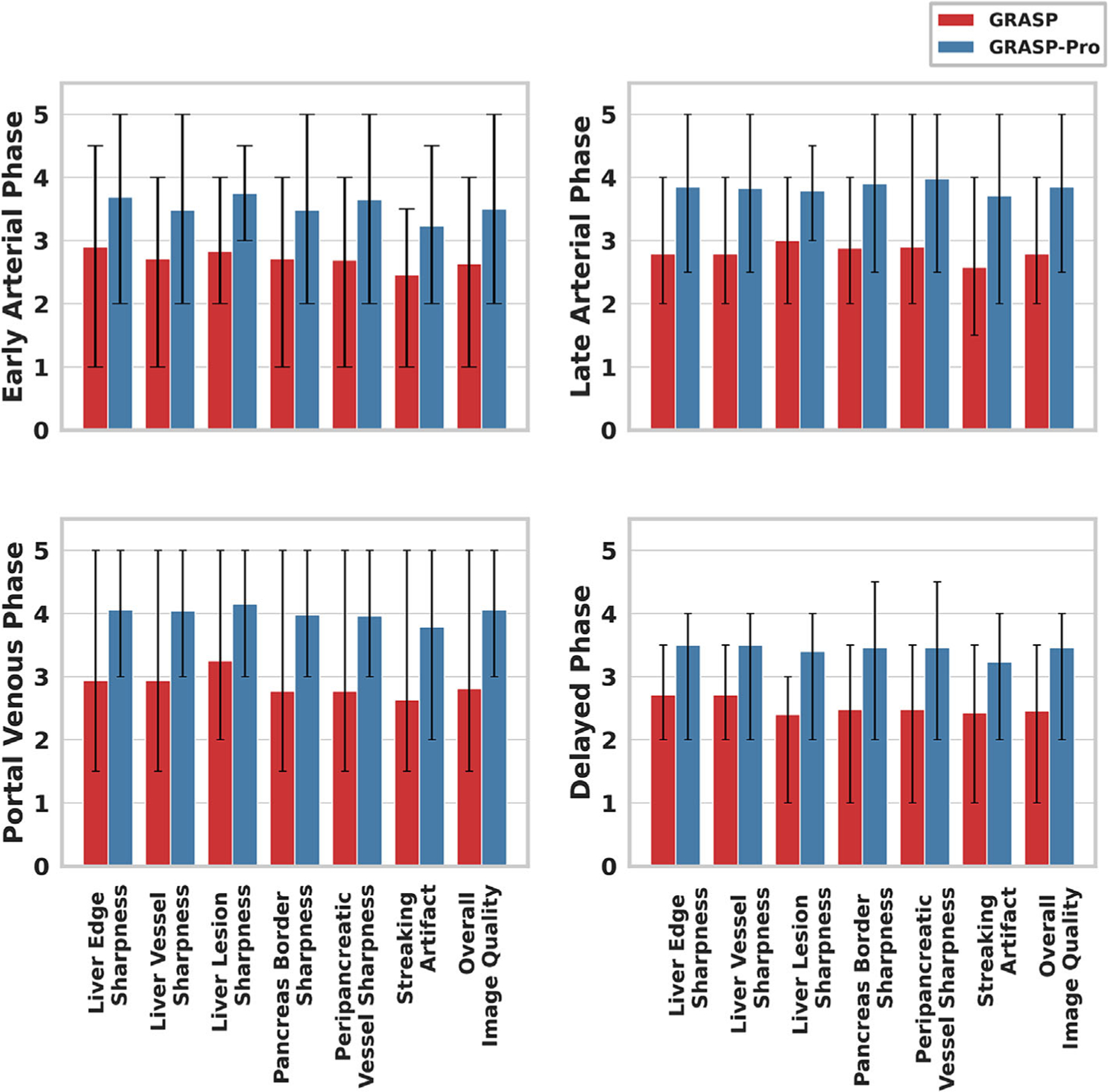
Respiratory motion-induced image blurring and artifacts can compromise image quality in dynamic contrast-enhanced MRI (DCE-MRI) of the liver. Despite remarkable advances in respiratory motion detection and compensation in past years, these techniques have not yet seen widespread clinical adoption. The accuracy of image-based motion detection can be especially compromised in the presence of contrast enhancement and/or in situations involving deep and/or irregular breathing patterns. This work proposes a framework that combines GRASP-Pro (Golden-angle RAdial Sparse Parallel MRI with imProved performance) MRI with a new radial sampling scheme called navi-stack-of-stars for free-breathing DCE-MRI of the liver without the need for explicit respiratory motion compensation. A prototype 3D golden-angle radial sequence with a navi-stack-of-stars sampling scheme that intermittently acquires a 2D navigator was implemented. Free-breathing DCE-MRI of the liver was conducted in 24 subjects at 3T including 17 volunteers and 7 patients. GRASP-Pro reconstruction was performed with a temporal resolution of 0.34-0.45 s per volume, whereas standard GRASP reconstruction was performed with a temporal resolution of 15 s per volume. Motion compensation was not performed in all image reconstruction tasks. Liver images in different contrast phases from both GRASP and GRASP-Pro reconstructions were visually scored by two experienced abdominal radiologists for comparison. The nonparametric paired two-tailed Wilcoxon signed-rank test was used to compare image quality scores, and the Cohen's kappa coefficient was calculated to evaluate the inter-reader agreement. GRASP-Pro MRI with sub-second temporal resolution consistently received significantly higher image quality scores (P < 0.05) than standard GRASP MRI throughout all contrast enhancement phases and across all assessment categories. There was a substantial inter-reader agreement for all assessment categories (ranging from 0.67 to 0.89). The proposed technique using GRASP-Pro reconstruction with navi-stack-of-stars sampling holds great promise for free-breathing DCE-MRI of the liver without respiratory motion compensation.

Keywords: DCE‐MRI; GRASP‐Pro; free‐breathing imaging; motion compensation; navi‐stack‐of‐star; radial sampling.


Conflict of interest statement

CONFLICT OF INTEREST STATEMENT

Dr. Feng and Dr. Chandarana are co-inventors of a patent on the GRASP MRI technique.

Figures

FIGURE 1
FIGURE 1
Diagram of navi-stack-of-stars sampling. Based on golden angle stack-of-star sampling, the rotation angle is set to be 0 degree every M spokes (here for illustration, M=2). The 2D FFT of stacks of 0 degree serve as navigators to track motion and contrast change. The adjacent spokes of each 2D navigator are grouped together to recover one imaging frame.
FIGURE 2
FIGURE 2
(A) Comparison of GRASP and GRASP-Pro reconstructed dynamic images for a healthy volunteer. The temporal resolution for GRASP reconstruction is 15 s per volume, whereas the GRASP-Pro reconstruction can achieve a temporal resolution of 0.5 s per volume. The GRASP-Pro reconstruction with higher temporal resolution shows less temporal blurring and is able to recover more details of structure at the arterial phase of contrast enhancement. (B) The contrast enhancement curves from GRASP, GRASP-Pro, and NUFF reconstruction methods. The NUFFT method does not impose temporal regularization; therefore, its temporal dynamics is used here as a reference curve.
FIGURE 3
FIGURE 3
(A) Dynamics images of an axial slice using GRASP and GRASP-Pro reconstruction for a healthy volunteer. The GRASP reconstruction has a lower temporal resolution and shows motion blurring (green arrow), whereas the GRASP-Pro reconstruction preserves distinctive structures with higher temporal resolutions. (B) Dynamics images of a coronal slice using GRASP and GRASP-Pro reconstruction for a healthy volunteer. The GRASP reconstruction shows visible motion-induced artifact, especially at the hepatic dome region where the displacement from respiratory motion is large.
FIGURE 4
FIGURE 4
Comparison of GRASP and GRASP-Pro reconstruction in a clinically confirmed HCC patient (HCC patient 1). (A) The liver lesions are clearly delineated in dynamic images of four contrast enhancement phases using GRASP-Pro reconstruction. The GRASP-reconstructed dynamics images contain motion blurring making the liver lesions less distinctive visually. (B) The contrast enhancement curves of GRASP and GRASP-Pro reconstruction align well for the abdominal aorta, liver parenchyma, and HCC regions. GRASP-Pro has a higher temporal resolution, and it captures more detailed dynamic contrast changes compared with GRASP-reconstructed dynamic images.
FIGURE 5
FIGURE 5
Comparison of GRASP and GRASP-Pro reconstruction of one coronal slice and one reformatted sagittal slice for the HCC patient 1. Motion-induced artifacts are prominent in GRASP reconstruction, especially at the superior half of the liver.
FIGURE 6
FIGURE 6
Comparison of conventional GRASP, NUFFT, GRASP, and GRASP-Pro reconstruction at a high temporal resolution of another image slice in the same HCC patient as shown in Figure 5 (HCC patient 1). GRASP-Pro-reconstructed images show a clearer definition of liver lesions and anatomical structures compared with the conventional GRASP reconstruction. When the temporal resolution NUFFT and GRASP reconstruction matches the proposed GRASP-Pro reconstruction, the image becomes unreadable.
FIGURE 7
FIGURE 7
Comparison of GRASP with GRASP-Pro in HCC patient 2 and patient 4 for representative image slices. (A) The tumor and the liver vessels presenting (pointed by the yellow arrows) are better delineated in the GRASP-Pro-reconstructed images. (B) The patient previously underwent liver resection, and the region of the surgery is clearly outlined using GRASP-Pro reconstruction even during the early arterial phase of the contrast enhancement.
FIGURE 8
FIGURE 8
Readers’ scores for image quality assessments of each contrast enhancement phase. Scores of two radiologists are averaged to calculate mean, max, and min values for each category for all 24 subjects. The error bar on the bar plot represents the max and min values. The mean scores for GRASP-Pro images are consistently higher than that of GRASP images. The scores of the delayed phase have the lowest range for the scores compared with other DCE phases.
FIGURE 9
FIGURE 9
Comparison of GRASP-Pro reconstruction using three basis estimation schemes for an HCC patient. Images of the early arterial phase are shown on the left, and the temporal x-t profiles of a line crossing the right lobe of the liver are shown on the right. The lesions are clearly delineated in the image reconstructed using navigator-based basis estimation, whereas strong artifacts are present when using GRASP-Pro with basis estimated from the low-resolution images and from the k-space centers. The x-t profile from navigator-based basis estimation captures the contrast uptake and the respiratory motion, whereas the temporal x-t profiles exhibit strong signal jittering and artifacts when using low-resolution image-based and k-space center basis estimation. Dynamic movies corresponding to this figure can be found in the supporting movie document, where the improvement with navigator-based basis estimation is more apparent.
FIGURE 10
FIGURE 10
Comparison of GRASP-Pro reconstruction using three basis estimation schemes for a healthy volunteer. Images of the early arterial phase from three versions of GRASP-Pro reconstruction methods are shown on the left alongside the temporal x-t profiles of a line crossing the right lobe of liver showing on the right. GRASP-Pro reconstruction with navigator-based basis estimation achieves superior quality, visible in a single image frame and more pronounced in the temporal x-t profiles. Dynamic movies corresponding to this figure can be found in the supporting movie document, where the improvement with navigator-based basis estimation is more apparent.
FIGURE 11
FIGURE 11
Dynamic image frames during respiratory cycle for two HCC patients. The time interval between frames is 0.34 s. This high temporal resolution allows for clear image reconstruction using GRASP-Pro without explicit motion compensation.

Similar articles

Cited by

References

    1. Elsayes KM, Narra VR, Yin Y, Mukundan G, Lammle M, Brown JJ. Focal hepatic lesions: diagnostic value of enhancement pattern approach with contrast-enhanced 3D gradient-echo MR imaging. Radiographics. 2005;25(5):1299–1320. doi:10.1148/rg.255045180 - DOI - PubMed
    1. Do RKG, Rusinek H, Taouli B. Dynamic contrast-enhanced MR imaging of the liver: current status and future directions. Magn Reson Imaging Clin N am 2009;17(2):339–349. doi:10.1016/j.mric.2009.01.009 - DOI - PubMed
    1. Quillin SP, Atilla S, Brown JJ, Borrello JA, Yu C-Y, Pilgram TK. Characterization of focal hepatic masses by dynamic contrast-enhanced MR imaging: findings in 311 lesions. Magn Reson Imaging. 1997;15(3):275–285. doi:10.1016/S0730-725X(96)00226-3 - DOI - PubMed
    1. Maki JH, Chenevert TL, Prince MR. The effects of incomplete breath-holding on 3D MR image quality. J Magn Reson Imaging. 1997;7(6):1132–1139. doi:10.1002/jmri.1880070628 - DOI - PubMed
    1. Feng L, Grimm R, Block KT, et al. Golden-angle radial sparse parallel MRI: combination of compressed sensing, parallel imaging, and golden-angle radial sampling for fast and flexible dynamic volumetric MRI. Magn Reson Med 2014;72(3):707–717. doi:10.1002/mrm.24980 - DOI - PMC - PubMed

LinkOut - more resources