Diagnostic Performance of CLEIA Versus FEIA for KL-6 Peripheral and Alveolar Concentrations in Fibrotic Interstitial Lung Diseases: A Multicentre Study
- PMID: 39323282
- PMCID: PMC11520937
- DOI: 10.1002/jcla.25108
Diagnostic Performance of CLEIA Versus FEIA for KL-6 Peripheral and Alveolar Concentrations in Fibrotic Interstitial Lung Diseases: A Multicentre Study
Abstract
Background: Interstitial lung diseases (ILD) is a group of lung disorders characterized by interstitial lung thickening due to inflammatory and fibrotic processes. Krebs von den Lungen-6 (KL-6) is a molecule secreted by damaged type II alveolar pneumocytes in the alveolar space. The goal of the present study was to compare two detection methods of KL-6 in both bronchoalveolar lavage (BAL) and serum from ILD patients at the moment of diagnosis.
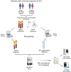
Methods: Patients with suspicious of ILD and followed at two Italian referral centres for rare lung diseases were included in the study. BAL fluid and serum were collected and analysed by chemiluminescent enzyme immunoassay (CLEIA) and fluorescent enzyme immunoassay (FEIA) methods provided by Tosoh Biosciences.
Results: A total of 158 (mean age ± standard deviation, 61.5 ± 13.7, 65 females) patients were enrolled. A total of, 36 had diagnosis of idiopathic pulmonary fibrosis (IPF), 74 sarcoidosis, 15 connective tissue disease-ILD (CTD-ILD) and 33 other ILD. Diagnostic agreement between two methods was demonstrated for both BAL (r = 0.707, p < 0.0001) and serum (r = 0.816, p < 0.0001). BAL KL-6 values were lower than serum (p < 0.0001). IPF patients had higher serum KL-6 concentration than other ILDs (p = 0.0294), while BAL KL-6 values were lower in IPF than in non-IPF (p = 0.0023).
Conclusion: This study explored KL-6 concentrations through the CLEIA method in serum and BAL of patients with various ILDs, showing significant differences of biomarkers concentrations between IPF and other non-IPF ILDs. Our findings are promising as they provided further knowledge concerning KL-6 expression across different ILDs and may suggest its utility in differential diagnosis.
Keywords: Krebs von den Lungen‐6; bronchoalveolar lavage; diagnosis; interstitial lung diseases; pulmonary fibrosis.
© 2024 The Author(s). Journal of Clinical Laboratory Analysis published by Wiley Periodicals LLC.
Conflict of interest statement
The authors declare no conflicts of interest.
Figures


References
-
- Behr J., “Approach to the Diagnosis of Interstitial Lung Disease,” Clinics in Chest Medicine 33, no. 1 (2012): 1–10. - PubMed
-
- Geiser T. and Guler S., “Diagnosis of ILD,” Therapeutische Umschau 73, no. 1 (2016): 7–10. - PubMed
-
- Travis W. D., Costabel U., Hansell D. M., et al., “An Official American Thoracic Society/European Respiratory Society Statement: Update of the International Multidisciplinary Classification of the Idiopathic Interstitial Pneumonias,” American Journal of Respiratory and Critical Care Medicine 188, no. 6 (2013): 733–748. - PMC - PubMed
Publication types
MeSH terms
Substances
LinkOut - more resources
Full Text Sources
Medical
Research Materials
Miscellaneous

