. 2024 Sep 11:15:1455439.
doi: 10.3389/fphar.2024.1455439. eCollection 2024.

DJ-1 regulates mitochondrial function and promotes retinal ganglion cell survival under high glucose-induced oxidative stress

Affiliations

DJ-1 regulates mitochondrial function and promotes retinal ganglion cell survival under high glucose-induced oxidative stress

Hanhan Peng et al. Front Pharmacol. .

Abstract

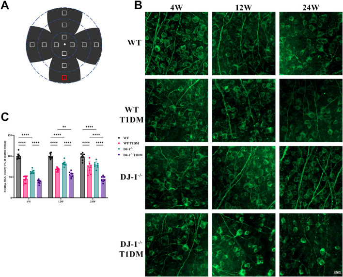
Purpose: This study aimed to investigate the antioxidative and neuroprotective effects of DJ-1 in mitigating retinal ganglion cell (RGC) damage induced by high glucose (HG).

Methods: A diabetic mouse model and an HG-induced R28 cell model were employed for loss- and gain-of-function experiments. The expression levels of apoptosis and oxidative stress-related factors, including Bax, Bcl-2, caspase3, Catalase, MnSOD, GCLC, Cyto c, and GPx-1/2, were assessed in both animal and cell models using Western blotting. Retinal structure and function were evaluated through HE staining, electroretinogram, and RGC counting. Mitochondrial function and apoptosis were determined using JC-1 and TUNEL staining, and reactive oxygen species (ROS) measurement.

Results: In the mouse model, hyperglycemia resulted in reduced retinal DJ-1 expression, retinal structural and functional damage, disrupted redox protein profiles, and mitochondrial dysfunction. Elevated glucose levels induced mitochondrial impairment, ROS generation, abnormal protein expression, and apoptosis in R28 cells. Augmenting DJ-1 expression demonstrated a restoration of mitochondrial homeostasis and alleviated diabetes-induced morphological and functional impairments both in vivo and in vitro.

Conclusion: This study provides novel insights into the regulatory role of DJ-1 in mitochondrial dynamics, suggesting a potential avenue for enhancing RGC survival in diabetic retinopathy.

Keywords: DJ-1; diabetic retinopathy; mitochondria; oxidative stress; retinal ganglion cells.


Conflict of interest statement

The authors declare that the research was conducted in the absence of any commercial or financial relationships that could be construed as a potential conflict of interest.

Figures

FIGURE 1
FIGURE 1
DJ-1 knockout exacerbates retinal structure impairment in diabetic mice. In wild-type or DJ-1−/− mice with or without diabetes 4, 12, and 24 weeks, the retinal structure was assessed by hematoxylin-eosin staining (n = 6 per group). Red arrows represent vacuolar changes, and blue arrows represent structural breaks or discontinuities. The scale bar represents 50 μm, with an enlarged inset highlighting the boxed area. GCL: ganglion cell layer; IPL: inner plexiform layer; INL: inner nuclear layer; OPL: outer plexiform layer; ONL: outer nuclear layer; IS: inner segments of photoreceptors.
FIGURE 2
FIGURE 2
DJ-1 knockout exacerbates retinal function impairment in diabetic mice. (A) Representative profile of ERG for wild-type and DJ-1−/− mice with or without diabetes; (B–D) Quantitative analysis of a-wave, b-wave, and OPs, respectively (n = 5 per group). OPs: oscillatory potentials. The comparison was based on two-way ANOVA analysis. P values of <0.05, <0.01, <0.001, and <0.0001 are indicated by *, **, ***, and ****, respectively.
FIGURE 3
FIGURE 3
DJ-1 knockout exacerbates RGC loss in diabetic mice. (A) A schematic diagram of retina whole mounting; (B) In wild-type or DJ-1−/− mice with or without diabetes 4, 12, and 24 weeks, β III Tubulin-positive cells in whole mounted retinas were assessed by observing flat-mounted retinas, scale bar 20 μm; (C) Quantitative analysis of RGC number (n = 8 per group). The comparison was based on two-way ANOVA analysis. P values of <0.01, and <0.0001 are indicated by **, and ****, respectively.
FIGURE 4
FIGURE 4
DJ-1 knockout exacerbates abnormalities in retinal protein expression. (A) Immunofluorescence staining showed the distribution of DJ-1 protein in the retina, scale bar 50 μm; (B) Western blotting of 12-week wild-type or DJ-1−/− mice with or without diabetes, including DJ-1, GCLC, caspase3, and GPx-1/2 (n = 3 per group); (C–F) Quantitative analysis of Western blot results for DJ-1, GCLC, caspase3, and GPx-1/2, respectively. GCL: ganglion cell layer; IPL: inner plexiform layer; INL: inner nuclear layer; OPL: outer plexiform layer; ONL: outer nuclear layer. The comparison was based on one-way ANOVA analysis. P values of <0.05, <0.01, <0.001, and <0.0001 are indicated by *, **, ***, and ****, respectively.
FIGURE 5
FIGURE 5
DJ-1 overexpression rescues diabetic abnormalities in the retinal structure and RGC apoptosis. (A) Hematoxylin-eosin staining showed retinal structure in mice with intravitreal injection of PARK7 overexpression virus and control virus (n = 5 per group). The scale bar represents 50 μm, with an enlarged inset highlighting the boxed area; (B) The β III Tubulin-positive cells in whole mounted retinas were assessed by observing flat-mounted retinas (n = 5 per group), scale bar 500 μm, the picture below is an enlargement of the one above, scale bar 50 μm; (C–D) Quantitative analysis of RGC number. RGC: retinal ganglion cell; GCL: ganglion cell layer; IPL: inner plexiform layer; INL: inner nuclear layer; OPL: outer plexiform layer; ONL: outer nuclear layer; IS/OS: inner segments of photoreceptors/outer segments of photoreceptors; NC: negative control; OE: overexpression. The comparison was based on an unpaired two-tailed t-test. P values of <0.01, and <0.0001 are indicated by ** and ****, respectively.
FIGURE 6
FIGURE 6
DJ-1 overexpression rescues diabetic retinal function and oxidative stress. (A) Representative profile of ERG for diabetic mice with DJ-1 NC and OE virus (n = 5 per group); (B) Western blotting was used to detect protein expression in diabetic mice with DJ-1 NC and OE virus, including DJ-1, Catalase, caspase3, and Cyto c (n = 3 per group); (C–E) Quantitative analysis of a-wave, b-wave, and OPs, respectively; (F) Quantitative analysis of Western blot results for DJ-1, Catalase, caspase3, and Cyto c. NC: negative control; OE: overexpression. The comparison was based on an unpaired two-tailed t-test. P values of <0.05, <0.01, <0.001, and <0.0001 are indicated by *, **, ***, and ****, respectively.
FIGURE 7
FIGURE 7
High glucose induces mitochondrial damage, apoptosis, and oxidative stress in R28 cells. (A) JC-1 staining showed mitochondrial transmembrane potential in R28 cells in normal control and high glucose treatment (n = 3 independent experiments), scale bar 100 μm; (B) Quantitative fluorescence density results of JC-1 ratio (poly/mono); (C) TUNEL staining showed R28 cell apoptosis (n = 3 independent experiments), scale bar 100 μm; (D) Quantitative analysis of apoptotic cell number results. (E) Western blotting results showed that high glucose challenge induced changes in DJ-1, GPx-1/2, MnSOD/SOD2, Catalase, Bcl-2, Bax, Bax/Bcl-2, and Cyto c protein expression (n = 3 independent experiments); (F–M) Quantitative analysis of Western blot results for DJ-1, GPx-1/2, MnSOD/SOD2, Catalase, Bcl-2, Bax, Bax/Bcl-2, and Cyto c, respectively. TUNEL: Terminal deoxynucleotidyl transferase dUTP nick end labeling. The comparison was based on an unpaired two-tailed t-test. P values of <0.05, <0.01, and <0.0001 are indicated by *, **, and ****, respectively.
FIGURE 8
FIGURE 8
DJ-1 regulates mitochondrial function, ROS generation, and cell apoptosis in R28 cells. (A) JC-1 staining showed mitochondrial transmembrane potential in R28 cells in control, high glucose, and high glucose with lentiviruses treatment (n = 3 independent experiments), scale bar 100 μm; (B) Quantitative fluorescence density results of JC-1 ratio (poly/mono); (C) ROS staining showed ROS generation in R28 cells in control, high glucose, and high glucose with lentiviruses treatment (n = 3 independent experiments); (D) Quantitative fluorescence density results of ROS generation; (E) TUNEL staining showed that DJ-1 overexpression significantly decreased the number of apoptotic R28 cells induced by high glucose (n = 3 independent experiments), scale bar 50 μm; (F) Quantitative analysis of apoptotic cell number results. ROS: reactive oxygen species; TUNEL: Terminal deoxynucleotidyl transferase dUTP nick end labeling; NC: negative control; OE: overexpression. The comparison was based on one-way ANOVA analysis. P values of <0.0001 are indicated by ****.
FIGURE 9
FIGURE 9
DJ-1 overexpression rescues HG-induced disturbances in redox and apoptotic protein expression. (A–E) Western blotting results showed that DJ-1 overexpression significantly reversed high glucose-induced protein expression abnormalities, including DJ-1, Catalase, Bcl-2, Cyto c, and caspase3 (n = 3 independent experiments); (F–J) Quantitative analysis of Western blot results for DJ-1, Catalase, Bcl-2, Cyto c, and caspase3, respectively. The comparison was based on one-way ANOVA analysis. P values of <0.05, <0.01, <0.001, and <0.0001 are indicated by *, **, ***, and ****, respectively.
FIGURE 10
FIGURE 10
DJ-1 enhances mitochondrial function and reduces oxidative stress, thereby promoting retinal ganglion cell survival in diabetic retinopathy.

Similar articles

Cited by

References

    1. Adebayo M., Singh S., Singh A. P., Dasgupta S. (2021). Mitochondrial fusion and fission: the fine-tune balance for cellular homeostasis. FASEB J. 35, e21620. 10.1096/fj.202100067R - DOI - PMC - PubMed
    1. Amato R., Catalani E., Dal Monte M., Cammalleri M., Cervia D., Casini G. (2022). Morpho-functional analysis of the early changes induced in retinal ganglion cells by the onset of diabetic retinopathy: the effects of a neuroprotective strategy. Pharmacol. Res. 185, 106516. 10.1016/j.phrs.2022.106516 - DOI - PubMed
    1. Amatullah H., Maron-Gutierrez T., Shan Y., Gupta S., Tsoporis J. N., Varkouhi A. K., et al. (2021). Protective function of DJ-1/PARK7 in lipopolysaccharide and ventilator-induced acute lung injury. Redox Biol. 38, 101796. 10.1016/j.redox.2020.101796 - DOI - PMC - PubMed
    1. Carrasco E., Hernandez C., Miralles A., Huguet P., Farres J., Simo R. (2007). Lower somatostatin expression is an early event in diabetic retinopathy and is associated with retinal neurodegeneration. Diabetes Care 30, 2902–2908. 10.2337/dc07-0332 - DOI - PubMed
    1. Cecilia O. M., Jose Alberto C. G., Jose N. P., Ernesto German C. M., Ana Karen L. C., Luis Miguel R. P., et al. (2019). Oxidative stress as the main target in diabetic retinopathy pathophysiology. J. Diabetes Res. 2019, 8562408. 10.1155/2019/8562408 - DOI - PMC - PubMed

LinkOut - more resources