

Comprehensive Classification System for Localized Alveolar Bone Deficiencies in Treatment Planning for Dental Implants: A Proposed Classification and Prevalence Study
- PMID: 39323713
- PMCID: PMC11422703
- DOI: 10.7759/cureus.67769
Comprehensive Classification System for Localized Alveolar Bone Deficiencies in Treatment Planning for Dental Implants: A Proposed Classification and Prevalence Study
Abstract
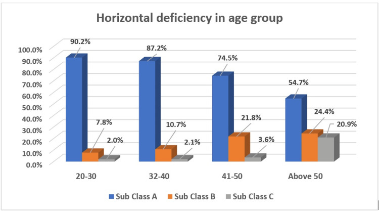
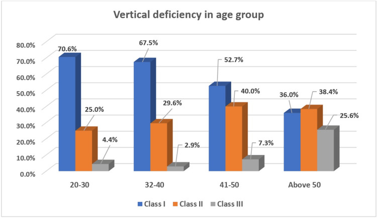
Introduction The current classification of alveolar bone defects remains ambiguous. This study aims to classify and evaluate the prevalence of bone deformities using a comprehensive classification system for localized alveolar bone deficiencies in dental implant treatment planning. Methods This cross-sectional prospective clinical trial included 698 participants (353 females and 345 males), patients with localized alveolar bone deficiencies. The clinical parameters evaluated were keratinized tissue width (KTW) and gingival thickness (GT) at the edentulous site. The width and height of alveolar bone deficiency at the site of implant placement were measured as horizontal deficiency (HD) and vertical deficiency (VD). Results Out of 698 patients, 566 (81.10%) had Subclass A horizontal deficiencies (HD), 99 (14.20%) had Subclass B HD, and 33 (4.70%) had Subclass C HD. Regarding vertical deficiencies (VD), 426 (61%) had Class I VD, 222 (31.80%) had Class II VD, and 50 (7.20%) had Class III VD. Younger individuals (20-30 years) predominantly exhibited Subclass A HD, whereas older participants (above 50 years) showed more severe deficiencies (Subclass B and C HD, and Class II and III VD). Gender analysis indicated no significant difference in HD prevalence but a significant difference in VD, with males more commonly presenting Class I VD and females exhibiting more Class II VD. Conclusion The study reveals significant associations between age and both HD and VD, indicating that older individuals tend to have more severe deficiencies. The study's findings underscore the importance of the proposed classification system in accurately identifying alveolar bone deficiencies and guiding appropriate treatment strategies, thereby improving clinical outcomes in dental implant therapy.
Keywords: alveolar ridge; atrophy; classification; dental implants; prevalence.
Copyright © 2024, Kolte et al.
Conflict of interest statement
Human subjects: Consent was obtained or waived by all participants in this study. Institutional Ethics Committee, VSPM (Vidya Shikshan Prasarak Mandal) Dental College and Research Centre, Nagpur issued approval IEC/VSPMDCRC/05/2022, dated January 15, 2022. Animal subjects: All authors have confirmed that this study did not involve animal subjects or tissue. Conflicts of interest: In compliance with the ICMJE uniform disclosure form, all authors declare the following: Payment/services info: All authors have declared that no financial support was received from any organization for the submitted work. Financial relationships: All authors have declared that they have no financial relationships at present or within the previous three years with any organizations that might have an interest in the submitted work. Other relationships: All authors have declared that there are no other relationships or activities that could appear to have influenced the submitted work.
Figures
Similar articles
-
The Prevalence of Alveolar Ridge Defects According to Seibert's Classification: A Cross-Sectional Study.Cureus. 2024 Dec 12;16(12):e75602. doi: 10.7759/cureus.75602. eCollection 2024 Dec. Cureus. 2024. PMID: 39803038 Free PMC article.
-
[The modified lip-tooth-ridge classification: a guide for edentulous maxillary arches].Hua Xi Kou Qiang Yi Xue Za Zhi. 2018 Jun 1;36(3):233-239. doi: 10.7518/hxkq.2018.03.001. Hua Xi Kou Qiang Yi Xue Za Zhi. 2018. PMID: 29984920 Free PMC article. Chinese.
-
The influence of initial alveolar ridge defect morphology on the outcome of implants in augmented atrophic posterior mandible: an exploratory retrospective study.Clin Oral Implants Res. 2017 Oct;28(10):e208-e217. doi: 10.1111/clr.12991. Epub 2016 Nov 2. Clin Oral Implants Res. 2017. PMID: 27804178
-
Interventions for replacing missing teeth: alveolar ridge preservation techniques for dental implant site development.Cochrane Database Syst Rev. 2021 Apr 26;4(4):CD010176. doi: 10.1002/14651858.CD010176.pub3. Cochrane Database Syst Rev. 2021. PMID: 33899930 Free PMC article.
-
The sandwich osteotomy technique to treat vertical alveolar bone defects prior to implant placement: a systematic review.Clin Oral Investig. 2020 Mar;24(3):1073-1089. doi: 10.1007/s00784-019-03183-6. Epub 2020 Jan 11. Clin Oral Investig. 2020. PMID: 31927693
References
-
- Bone healing and soft tissue contour changes following single-tooth extraction: a clinical and radiographic 12-month prospective study. Schropp L, Wenzel A, Kostopoulos L, Karring T. https://pubmed.ncbi.nlm.nih.gov/12956475/ Int J Periodontics Restorative Dent. 2003;23:313–323. - PubMed
-
- Hard-tissue alterations following immediate implant placement in extraction sites. Botticelli D, Berglundh T, Lindhe J. J Clin Periodontol. 2004;31:820–828. - PubMed
-
- Dynamics of bone tissue formation in tooth extraction sites: an experimental study in dogs. Cardaropoli G, Araújo M, Lindhe J. J Clin Periodontol. 2003;30:761–772. - PubMed
-
- Dimensional ridge alterations following tooth extraction. An experimental study in the dog. Araújo MG, Lindhe J. J Clin Periodontol. 2005;32:212–218. - PubMed
-
- Bone healing dynamics at buccal peri-implant sites. Qahash M, Susin C, Polimeni G, Hall J, Wikesjö UM. Clin Oral Implants Res. 2008;19:48–54. - PubMed
LinkOut - more resources
Full Text Sources
Miscellaneous