. 2024 Sep 18;5(11):1140-1146.
doi: 10.1039/d4cb00183d. Online ahead of print.

Rapid formation of N ε-(carboxymethyl)lysine (CML) from ribose depends on glyoxal production by oxidation

Affiliations

Rapid formation of N ε-(carboxymethyl)lysine (CML) from ribose depends on glyoxal production by oxidation

Hikari Sugawa et al. RSC Chem Biol. .

Abstract

N ε-(Carboxymethyl)lysine (CML) is a major advanced glycation end-product (AGE) involved in protein dysfunction and inflammation in vivo. Its accumulation increases with age and is enhanced with the pathogenesis of diabetic complications. Therefore, the pathways involved in CML formation should be elucidated to understand the pathological conditions involved in CML. Ribose is widely used in glycation research because it shows a high reactivity with proteins to form AGEs. We previously demonstrated that ribose generates CML more rapidly than other reducing sugars, such as glucose; however, the underlying mechanism remains unclear. In this study, we focused on the pathway of CML formation from ribose. As a result, glyoxal (GO) was the most abundant product generated from ribose among the tested reducing sugars and was significantly correlated with CML formation from ribose-modified protein. The coefficient of determination (R 2) for CML formation between the ribose-modified protein and Amadori products or the ribose degradation product (RDP)-modified protein was higher for the RDP-modified protein. CML formation from ribose degradation products (RDP) incubated with protein significantly correlated with CML formation from GO-modified protein (r s = 0.95, p = 0.0000000869). GO and CML formation were inhibited by diethylenetriaminepentaacetic acid (DTPA) and enhanced by iron chloride. Additionally, flavonoid compounds such as isoquercetin, which are known to inhibit CML, also inhibited GO formation from ribose and CML formation. In conclusion, ribose undergoes auto-oxidation and oxidative cleavage between C-2 and C-3 to generate GO and enhance CML accumulation.


Conflict of interest statement

There are no conflicts to declare.

Figures

Fig. 1
Fig. 1. Evaluation of CML formation derived from reducing sugars. CML formation was evaluated using ELISA with a monoclonal CML antibody. (A) This study aimed to evaluate the pathways involved in CML development. (B) Comparison of CML formation using various reducing sugars mixed with gelatine. Different superscripts indicate statistically significant differences between groups (P < 0.01). (C) CML formation on the Amadori rearrangement products derived from each reducing sugar. CML formation was evaluated via oxidation of each sugar with 0.4 mM anhydrous FeCl2 in the absence or presence of 5 mM H2O2. The different reducing sugars are indicated as follows: ribose: ▲, glucose: ■, galactose: ×, mannose: ●, and fructose: ◆. Data obtained before and after oxidation are indicated by open and closed symbols, respectively (** p < 0.01 vs. before oxidation). (D) RDP was prepared by incubating each reducing sugar solution with a mixture of gelatin. The different reducing sugars are indicated as follows: ribose: ▲, glucose: ■, galactose: ×, mannose: ●, and fructose: ◆. Different superscripts indicate statistically significant differences between groups (P < 0.01). All data are presented as the mean ± SD (n = 3).
Fig. 2
Fig. 2. GO formation from ribose. (A) Time course of CML formation in ribose gelatine. Different superscripts indicate statistically significant differences between the groups (p < 0.001). (B) Predicted structure of derivatized GO. The asterisk indicates carbon-13 in the internal standard. (C) GO formation was evaluated using LC-ESI-QTOF. Fragment pattern of derivatized GO. MS/MS spectra of GO standard (i), GO in ribose solution (ii), and GO in 13C1 ribose solution (iii). (D) GO formation in reducing sugar solutions. The different reducing sugars have been indicated as follows: ribose: ▲, glucose: ■, galactose: ×, mannose: ●, and fructose: ◆. (E) Extracted ion chromatogram (±0.01) of derivatized GO. Different superscripts indicate statistically significant differences between the groups (p < 0.001). All data are presented as the mean ± SD (n = 3).
Fig. 3
Fig. 3. Correlation of GO-derived CML with CML and GO formation from ribose. CML formation was evaluated using ELISA. The GO content was measured using LC-ESI-QTOF. (A) Correlation between CML formation on ribose–gelatin and GO content in the ribose solution (rs = 0.87, p = 0.0000275). (B) Correlation between CML formation on ribose–gelatin and CML formation on RDP–gelatin (R2 = 0.8378). (C) Correlation between CML formation on ribose–gelatin and CML formation on ribose-derived Amadori rearrangement products (R2 = 0.6871). (D) Time course of CML formation on the GO–gelatin. Different superscripts indicate statistically significant differences between groups (P < 0.01). (E) Correlation between CML formation on GO–gelatin and CML formation on RDP–gelatin (rs = 0.95, p = 0.0000000869).
Fig. 4
Fig. 4. Ribose-derived GO was produced in an oxidation-dependent manner. (A) To elucidate the mechanism of GO formation from ribose, 50 mM ribose was incubated with or without DTPA or FeCl3, followed by LC-ESI-QTOF analysis (n = 3). (B) A mixture of 30 mM ribose and gelatin was incubated with or without DTPA or FeCl3 followed by ELISA (n = 3). Data are presented as the mean ± SD. Different superscripts indicate statistically significant differences between groups (P < 0.01).
Fig. 5
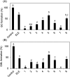
Fig. 5. Inhibitory effect of ELE-related compounds on GO and CML formation. Ribose was incubated with/without (control) the ELE-related compounds. (A) Inhibitory effect on GO formation was evaluated using LC-ESI-QTOF. Gelatin and ribose were incubated in the presence or absence (control) of the ELE-related compounds. (B) The inhibitory effect on CML formation was then evaluated using ELISA (absorbance 492 nm). Data are expressed as formation (%) relative to that of the control (without ELE-related compounds). The ELE related compounds used are shown in Table 1: 1, isoquercetin; 2, rutin; 3, quercetin-3-O-sambubioside; 4, astragalin; 5, kaempferol-3-O-rutinoside; 6, kaempferol-3-O-(6′′-acetyl)-glucoside; 7, quercetin; 8, kaempferol. Data are presented as mean ± SD (n = 3). Different superscripts indicate statistically significant differences between groups (p < 0.05).
Fig. 6
Fig. 6. Proposed mechanism of GO formation from ribose.

Similar articles

Cited by

References

    1. Nagai R. Shirakawa J. Fujiwara Y. Ohno R. Moroishi N. Sakata N. Nagai M. J. Clin. Biochem. Nutr. 2014;55:1–6. doi: 10.3164/jcbn.13-112. doi: 10.3164/jcbn.13-112. - DOI - DOI - PMC - PubMed
    1. Reddy S. Bichler J. Wells-Knecht K. J. Thorpe S. R. Baynes J. W. Biochemistry. 1995;34:10872–10878. doi: 10.1021/bi00034a021. doi: 10.1021/bi00034a021. - DOI - DOI - PubMed
    1. Dunn J. A. McCance D. R. Thorpe S. R. Lyons T. J. Baynes J. W. Biochemistry. 1991;30:1205–1210. doi: 10.1021/bi00219a007. doi: 10.1021/bi00219a007. - DOI - DOI - PubMed
    1. Sveen K. A. Karimé B. Jørum E. Mellgren S. I. Fagerland M. W. Monnier V. M. Dahl-Jørgensen K. Hanssen K. F. Diabetes Care. 2013;36:3712–3717. doi: 10.2337/dc13-0788. doi: 10.2337/dc13-0788. - DOI - DOI - PMC - PubMed
    1. Kralev S. Zimmerer E. Brueckmann M. Lang S. Kälsch T. Rippert A. Lin J. Borggrefe M. Hammes H. P. Süselbeck T. Clin. Chem. Lab. Med. 2009;47:446–451. doi: 10.1515/CCLM.2009.100. - DOI - PubMed