Cross-linking manipulation of waterborne biodegradable polyurethane for constructing mechanically adaptable tissue engineering scaffolds
- PMID: 39323747
- PMCID: PMC11422185
- DOI: 10.1093/rb/rbae111
Cross-linking manipulation of waterborne biodegradable polyurethane for constructing mechanically adaptable tissue engineering scaffolds
Abstract
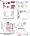
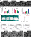
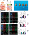
Mechanical adaptation of tissue engineering scaffolds is critically important since natural tissue regeneration is highly regulated by mechanical signals. Herein, we report a facile and convenient strategy to tune the modulus of waterborne biodegradable polyurethanes (WBPU) via cross-linking manipulation of phase separation and water infiltration for constructing mechanically adaptable tissue engineering scaffolds. Amorphous aliphatic polycarbonate and trifunctional trimethylolpropane were introduced to polycaprolactone-based WBPUs to interrupt interchain hydrogen bonds in the polymer segments and suppress microphase separation, inhibiting the crystallization process and enhancing covalent cross-linking. Intriguingly, as the crosslinking density of WBPU increases and the extent of microphase separation decreases, the material exhibits a surprisingly soft modulus and enhanced water infiltration. Based on this strategy, we constructed WBPU scaffolds with a tunable modulus to adapt various cells for tissue regeneration and regulate the immune response. As a representative application of brain tissue regeneration model in vivo, it was demonstrated that the mechanically adaptable WBPU scaffolds can guide the migration and differentiation of endogenous neural progenitor cells into mature neurons and neuronal neurites and regulate immunostimulation with low inflammation. Therefore, the proposed strategy of tuning the modulus of WBPU can inspire the development of novel mechanically adaptable biomaterials, which has very broad application value.
Keywords: central nervous repair; mechanical adaptation; modulus; tissue engineering scaffold; waterborne polyurethane.
© The Author(s) 2024. Published by Oxford University Press.
Figures


Similar articles
-
A novel waterborne polyurethane with biodegradability and high flexibility for 3D printing.Biofabrication. 2020 May 12;12(3):035015. doi: 10.1088/1758-5090/ab7de0. Biofabrication. 2020. PMID: 32150742
-
Low-Initial-Modulus Biodegradable Polyurethane Elastomers for Soft Tissue Regeneration.ACS Appl Mater Interfaces. 2017 Jan 25;9(3):2169-2180. doi: 10.1021/acsami.6b15009. Epub 2017 Jan 12. ACS Appl Mater Interfaces. 2017. PMID: 28036169 Free PMC article.
-
Quantitative grafting of peptide onto the nontoxic biodegradable waterborne polyurethanes to fabricate peptide modified scaffold for soft tissue engineering.J Mater Sci Mater Med. 2011 Apr;22(4):819-27. doi: 10.1007/s10856-011-4265-z. Epub 2011 Mar 1. J Mater Sci Mater Med. 2011. PMID: 21360121
-
Recent advances in tissue engineering scaffolds based on polyurethane and modified polyurethane.Mater Sci Eng C Mater Biol Appl. 2021 Jan;118:111228. doi: 10.1016/j.msec.2020.111228. Epub 2020 Aug 8. Mater Sci Eng C Mater Biol Appl. 2021. PMID: 33254956 Review.
-
Biodegradable polyurethane scaffolds in regenerative medicine: Clinical translation review.J Biomed Mater Res A. 2022 Aug;110(8):1460-1487. doi: 10.1002/jbm.a.37394. Epub 2022 Apr 28. J Biomed Mater Res A. 2022. PMID: 35481723 Review.
References
-
- Ma Y, Lin M, Huang G, Li Y, Wang S, Bai G, Lu T, Xu F.. 3D spatiotemporal mechanical microenvironment: a hydrogel-based platform for guiding stem cell fate. Adv Mater 2018;30:1705911. - PubMed
-
- Wang S, Zhu C, Zhang B, Hu J, Xu J, Xue C, Bao S, Gu X, Ding F, Yang Y, Gu X, Gu Y.. BMSC-derived extracellular matrix better optimizes the microenvironment to support nerve regeneration. Biomaterials 2022;280:121251. - PubMed
-
- Li Y, Xiao Y, Liu C.. The horizon of materiobiology: a perspective on material-guided cell behaviors and tissue engineering. Chem Rev 2017;117:4376–421. - PubMed
-
- Ye K, Wang X, Cao L, Li S, Li Z, Yu L, Ding J.. Matrix stiffness and nanoscale spatial organization of cell-adhesive ligands direct stem cell fate. Nano Lett 2015;15:4720–9. - PubMed
-
- Wang Y, Liang R, Lin J, Chen J, Zhang Q, Li J, Wang M, Hui X, Tan H, Fu Q.. Biodegradable polyurethane nerve guide conduits with different moduli influence axon regeneration in transected peripheral nerve injury. J Mater Chem B 2021;9:7979–90. - PubMed
LinkOut - more resources
Full Text Sources
Research Materials

