Review
. 2024 Sep 11:15:1446072.
doi: 10.3389/fimmu.2024.1446072. eCollection 2024.

Role of cellular effectors in the induction and maintenance of IgA responses leading to protective immunity against enteric bacterial pathogens

Affiliations
Review

Role of cellular effectors in the induction and maintenance of IgA responses leading to protective immunity against enteric bacterial pathogens

Laura E Carreto-Binaghi et al. Front Immunol. .

Abstract

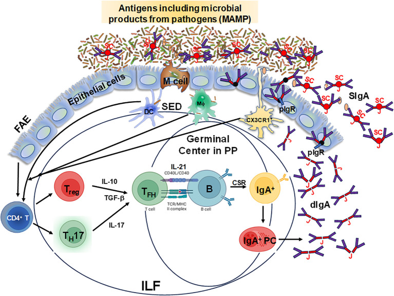
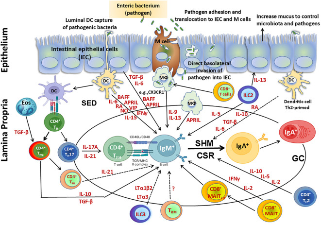
The mucosal immune system is a critical first line of defense to infectious diseases, as many pathogens enter the body through mucosal surfaces, disrupting the balanced interactions between mucosal cells, secretory molecules, and microbiota in this challenging microenvironment. The mucosal immune system comprises of a complex and integrated network that includes the gut-associated lymphoid tissues (GALT). One of its primary responses to microbes is the secretion of IgA, whose role in the mucosa is vital for preventing pathogen colonization, invasion and spread. The mechanisms involved in these key responses include neutralization of pathogens, immune exclusion, immune modulation, and cross-protection. The generation and maintenance of high affinity IgA responses require a delicate balance of multiple components, including B and T cell interactions, innate cells, the cytokine milieu (e.g., IL-21, IL-10, TGF-β), and other factors essential for intestinal homeostasis, including the gut microbiota. In this review, we will discuss the main cellular components (e.g., T cells, innate lymphoid cells, dendritic cells) in the gut microenvironment as mediators of important effector responses and as critical players in supporting B cells in eliciting and maintaining IgA production, particularly in the context of enteric infections and vaccination in humans. Understanding the mechanisms of humoral and cellular components in protection could guide and accelerate the development of more effective mucosal vaccines and therapeutic interventions to efficiently combat mucosal infections.

Keywords: IgA; T cells; enteric diseases; long term immune response; vaccination and controlled human infection model (CHIM).


Conflict of interest statement

The authors declare that the research was conducted in the absence of any commercial or financial relationships that could be construed as a potential conflict of interest.

Figures

Figure 1
Figure 1
Schematic representation of specialized follicle-associated epithelium in the intestinal lamina propria. Microfold (M) cells sample different types of antigens, including microbe-associated molecular patterns (MAMP), which move into the subepithelial dome (SED) and interact with dendritic cells (DC), T and B cells for the efficient induction of IgA responses from plasma cells leading to the production of dimeric IgA (dIgA) which is linked by the J chain (J). This molecule is picked up by the polymeric Ig receptor (pIgR) at the basal surface of the epithelial cells, transported and secreted from their apical surface into the lumen after cleavage of the pIgR leaving the secretory component (SC) resulting in secretory IgA (SIgA). Depicted is the follicle-associated epithelium (FAE) and the Peyer’s patches (PP) germinal centers and the isolated lymphoid follicles (ILF), where these cell interactions occur and are considered to be the primary IgA inductive sites within the gut-associated lymphoid tissue.
Figure 2
Figure 2
Cellular components involved directly or indirectly in the production of IgA. Secretory IgA (SIgA), representing at least 70% of all Ig produced in mammals, plays a major role as the first line of defense against adherence and invasion by enteric pathogens and neutralization of their toxins. SIgA is produced by IgA+ plasma cells residing in the intestinal lamina propria. Following infection with enteric pathogens, dendritic cells (DC) can either sample directly from the lumen or indirectly from the subepithelial dome (SED) resulting in their activation. Activated DC can directly influence class switch recombination (CSR) of IgM+ B cells by secretion of various molecules including cytokines (e.g., interleukin (IL)-6, interferon (IFN)γ, IL-15), B-cell-activating factor (BAFF), a proliferation-inducing ligand (APRIL), retinoic acid (RA), nitric oxide (NO) and vasoactive intestinal peptide (VIP). Activated DC can prime CD4+ T helper cells to differentiate into TH17, Treg, T follicular helper (TFH) and produce cytokines such as IL-17A, transforming growth factor (TGF) β, and IL-10, which are key contributors in supporting CSR. Eosinophils produce enzymes that activate TGF-β in the lamina propria, enhancing Treg control over the number of IgA+ plasma cells and also promote IgA CSR in the Peyer’s patches (PP). Following their differentiation from TH17 cells, PP TFH contribute to the production of cytokines (e.g., IL-17 and IL-21), while following their differentiation from Foxp3+ CD4+ T cells, T-Follicular regulatory cells (TFR) do not appear to support IgA switching through canonical TGF-β production but through IL-21. This process promotes a balanced development of TFH and TFR cells in PP. Treg interact with B cells directly or indirectly through cytokines (e.g., TGF-β, IL-10) and modulate their responses. In addition, Treg contribute to the germinal center reactions to promote secretion of IgA through the production of TGF-β, which is involved in IgA class switching. In addition, tissue resident memory (TRM) CD4+ T cells in PP may be important to rapidly replenish the plasma cell repertoire and for the strong memory IgA B cell responses elicited after re-exposure to oral antigens. CD8+ T cells interact with antigen presenting cells (APC) such as CX3CR1+ macrophages (MФ), promoting IgA production by B cells via secretion of IL-9 and IL-13. MAIT cells induce antibody production and B cell differentiation via cytokines such as IFNγ. Additionally, a subset of MAIT cells expressing CXCR5, TFH–like MAIT cells (MAITFH) directly provide B cell help. Furthermore, innate lymphoid cells (ILC) group 3 limit TFH responses and B cell class switching to IgA responses against commensal and pathogenic bacteria by production of lymphotoxins (LT) (e.g., LTα3, LTα1bβ). In contrast, ILC2 produce IL-13 which induce epithelial cells to produce mucus, controlling the microbiota and pathogens and impacting indirectly the regulation of IgA responses. Additionally, ILC2 secrete IL-5 which increase the production of IgA. Finally, intestinal epithelial cells (IEC) can produce cytokines (e.g., TGF-β, IL-6) and B cell factors such as BAFF and APRIL which sustain CSR leading to IgA B cells. Terminal differentiation into polymeric IgA (pIgA)-secreting plasma cells occurs in the lamina propria in a process regulated by cytokines and mediators secreted by activated CD4+ T helper 2 (TH2) cells (e.g., IL-2, IL-5, and IL-10) and DC (e.g., RA, IL-10, TGF-β, IL-6).

Similar articles

Cited by

References

    1. Pinheiro A, de Sousa-Pereira P, Strive T, Knight KL, Woof JM, Esteves PJ, et al. . Identification of a new European rabbit IgA with a serine-rich hinge region. PloS One. (2018) 13:e0201567. doi: 10.1371/journal.pone.0201567 - DOI - PMC - PubMed
    1. Flanagan JG, Rabbitts TH. Arrangement of human immunoglobulin heavy chain constant region genes implies evolutionary duplication of a segment containing gamma, epsilon and alpha genes. Nature. (1982) 300:709–13. doi: 10.1038/300709a0 - DOI - PubMed
    1. Lin M, Du L, Brandtzaeg P, Pan-Hammarstrom Q. IgA subclass switch recombination in human mucosal and systemic immune compartments. Mucosal Immunol. (2014) 7:511–20. doi: 10.1038/mi.2013.68 - DOI - PubMed
    1. Pakkanen SH, Kantele JM, Moldoveanu Z, Hedges S, Hakkinen M, Mestecky J, et al. . Expression of homing receptors on IgA1 and IgA2 plasmablasts in blood reflects differential distribution of IgA1 and IgA2 in various body fluids. Clin Vaccine immunol: CVI. (2010) 17:393–401. doi: 10.1128/CVI.00475-09 - DOI - PMC - PubMed
    1. Steffen U, Koeleman CA, Sokolova MV, Bang H, Kleyer A, Rech J, et al. . IgA subclasses have different effector functions associated with distinct glycosylation profiles. Nat Commun. (2020) 11:120. doi: 10.1038/s41467-019-13992-8 - DOI - PMC - PubMed

LinkOut - more resources