Review
. 2025 Feb;80(2):395-407.
doi: 10.1111/all.16337. Epub 2024 Sep 26.

How type-2 dendritic cells induce Th2 differentiation: Instruction, repression, or fostering T cell-T cell communication?

Affiliations
Review

How type-2 dendritic cells induce Th2 differentiation: Instruction, repression, or fostering T cell-T cell communication?

Franca Ronchese et al. Allergy. 2025 Feb.

Abstract

Allergic disease is caused by the activation of allergen-specific CD4+ type-2 T follicular helper cells (Tfh2) and T helper 2 (Th2) effector cells that secrete the cytokines IL-4, IL-5, IL-9, and IL-13 upon allergen encounter, thereby inducing IgE production by B cells and tissue inflammation. While it is accepted that the priming and differentiation of naïve CD4+ T cells into Th2 requires allergen presentation by type 2 dendritic cells (DC2s), the underlying signals remain unidentified. In this review we focus on the interaction between allergen-presenting DC2s and naïve CD4+ T cells in lymph node (LN), and the potential mechanisms by which DC2s might instruct Th2 differentiation. We outline recent advances in characterizing DC2 development and heterogeneity. We review mechanisms of allergen sensing and current proposed mechanisms of Th2 differentiation, with specific consideration of the role of DC2s and how they might contribute to each mechanism. Finally, we assess recent publications reporting a detailed analysis of DC-T cell interactions in LNs and how they support Th2 differentiation. Together, these studies are starting to shape our understanding of this key initial step of the allergic immune response.

Keywords: ILC2; Th2 differentiation; alarmins; allergen; dendritic cells.


Conflict of interest statement

FR, GW, SO, OL and MB declare no conflict of interest.

Figures

FIGURE 1
FIGURE 1
The allergic immune response. (A) During first exposure to allergen, naïve CD4+ T cells in lymph node become activated through contact with allergen‐presenting DC2s, and differentiate into type‐2 T follicular helper cells (Tfh2) or effector Th2 cells (Th2). (B) Upon allergen re‐exposure, Tfh2 in B cell follicles secrete IL‐4 to induce B cells to switch to IgE. Effector Th2 cells in non‐lymphoid tissues secrete cytokines including IL‐5, IL‐9 and IL‐13 to induce eosinophil and mast cell accumulation in the inflamed tissue, fibroblast proliferation with collagen deposition, and mucus production in the airway and intestinal tract. Together, these cytokines mediate the main features of allergic inflammation. Created with BioRender.com.
FIGURE 2
FIGURE 2
Dendritic cell (DC) differentiation and development. (A) The differentiation into conventional DCs (cDCs) subsets and DC3s occurs in the bone marrow where lymphoid progenitors (LPs), monocyte‐DC progenitors (MDPs) and common DC precursors (CDPs) give rise to pDC2‐like and cDC2 precursors (pre‐cDC2s) that can each differentiate into either cDC2As or cDC2Bs; pre‐cDC1s that differentiate into cDC1s; and DC3 progenitors (pro‐DC3s) that differentiate into DC3s. The capacity of each of these precursor or progenitor populations to establish resident and migratory DC populations has not been fully characterized. (B) Pre‐cDCs in bone marrow downregulate CXCR4 expression during maturation before being released into the bloodstream. From blood, pre‐cDCs enter LNs or spleen (not shown) to mature into the resident (res)‐cDC population. Alternatively, pre‐cDCs enter non‐lymphoid tissues to form the migratory cDC population which then migrates to draining LNs via lymph in a CCR7‐dependent manner. Created with BioRender.com.
FIGURE 3
FIGURE 3
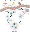
Allergen sensing by dendritic cells (DCs). (A) Metabolites produced by intestinal parasites stimulate tuft cells to produce IL‐25, which activates mig‐DC2s by inducing IL‐13 release by intestinal type‐2 innate lymphoid cells (ILC2). (B) Components of common allergens can signal through surface receptors including endocytic receptors, C‐type lectin receptors or Pattern‐recognition receptors to induce release of IL‐33, IL‐25 or Thymic stromal lymphopoietin (TSLP) by epithelial cells, and direct or indirect mig‐DC2 activation. (C) Protease allergens can interact with Protease‐activated receptors (PARs) on epithelial cells to induce the release of IL‐33, IL‐25 or TSLP to activate mig‐DCs either directly or indirectly. (D) Proteases in insect stings trigger pain and itch responses by triggering PARs on sensory neurons, with release of neuropeptides including substance P resulting in mig‐DC2 migration and activation. In all cases, direct or indirect (eg via ILC2‐derived IL‐13) mig‐DC2 activation by alarmins coincides with allergen uptake, migration to the draining lymph node and cognate interaction with naïve CD4+ T cells for Th2 differentiation. Created with BioRender.com.
FIGURE 4
FIGURE 4
Dermal type‐2 dendritic cells (DC2s) are innately programmed to induce Th2 differentiation without requiring activation of allergen sensing mechanisms. Dermal type‐2 innate lymphoid cells (ILC2s) in non‐inflamed, steady‐state skin produce IL‐13 without requiring the engagement of allergen sensing mechanisms and alarmin signals that are necessary in other tissues such as lung and small intestine, as detailed in Figure 3. Consequently, a dermal mig‐DC2 population expressing an IL‐4/IL‐13 transcriptional signature can be demonstrated in healthy human and mouse skin at steady state. These IL‐13‐conditioned mig‐DC2s express a heightened capacity to induce Th2 differentiation in lymph nodes. Created with BioRender.com.
FIGURE 5
FIGURE 5
DC2 signals for Th2 differentiation. (A) IL‐13 signaling can program mig‐DC2s to induce Th2 differentiation. Signaling by alarmins including TSLP, IL‐33 and IL‐25 can similarly program mig‐DC2s to Th2 differentiation, although the contribution of ILC2‐derived IL‐13 in each case has not been determined. (B) Reduced expression and signaling by pattern recognition receptors (PRRs) prevents DC2 expression of pro‐inflammatory cytokines such as IL‐12 and IL‐23, which drive Th1 and Th17 responses, respectively. (C) DC2s might express instructive cytokine or co‐stimulatory signals that direct CD4+ T cells to Th2 differentiation; to date, these signals have not been identified. (D) Weak T cell receptor (TCR) signaling through reduced processing and presentation of cognate antigen by DC2s, or impaired formation and duration of TCR synapse, could lead to Ca2+‐dependent signaling that is sufficient for Th2 but not Th1 activation. (E) Prolonged close contact between cDC2s and CD4+ T cells favors localized exchange of IL‐2 and IL‐4 cytokine signals which are necessary for the differentiation of naïve CD4+ T cells into effector Th2. Created with BioRender.com.

References

    1. Alladina J, Smith NP, Kooistra T, et al. A human model of asthma exacerbation reveals transcriptional programs and cell circuits specific to allergic asthma. Sci Immunol. 2023;8(83):eabq6352. - PMC - PubMed
    1. Ding Z, Mulder J, Robinson MJ. The origins and longevity of IgE responses as indicated by serological and cellular studies in mice and humans. Allergy. 2023;78(12):3103‐3117. - PMC - PubMed
    1. Kratchmarov R, Djeddi S, Dunlap G, et al. TCF1‐LEF1 co‐expression identifies a multipotent progenitor cell (T(H)2‐MPP) across human allergic diseases. Nat Immunol. 2024;25:902‐915. - PMC - PubMed
    1. Jutel M, Agache I, Zemelka‐Wiacek M, et al. Nomenclature of allergic diseases and hypersensitivity reactions: adapted to modern needs: an EAACI position paper. Allergy. 2023;78(11):2851‐2874. - PubMed
    1. Rothenberg‐Lausell C, Bar J, Dahabreh D, Renert‐Yuval Y, Del Duca E, Guttman‐Yassky E. Biologic and small molecule therapy for treating moderate to severe atopic dermatitis: mechanistic considerations. J Allergy Clin Immunol. 2024;154:20‐30. - PubMed