. 2024 Oct 15;137(20):jcs262198.
doi: 10.1242/jcs.262198. Epub 2024 Oct 23.

Expanding the field of view - a simple approach for interactive visualisation of electron microscopy data

Affiliations

Expanding the field of view - a simple approach for interactive visualisation of electron microscopy data

Jens Wohlmann. J Cell Sci. .

Abstract

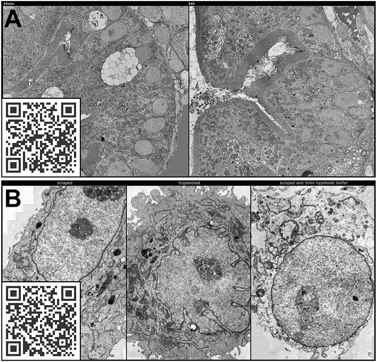
The unparalleled resolving power of electron microscopy is both a blessing and a curse. At 30,000× magnification, 1 µm corresponds to 3 cm in the image and the field of view is only a few micrometres or less, resulting in an inevitable reduction in the spatial data available in an image. Consequently, the gain in resolution is at the cost of loss of the contextual 'reference space', which is crucial for understanding the embedded structures of interest. This problem is particularly pronounced in immunoelectron microscopy, where the detection of a gold particle is crucial for the localisation of specific molecules. The common solution of presenting high-magnification and overview images side by side often insufficiently represents the cellular environment. To address these limitations, we propose here an interactive visualization strategy inspired by digital maps and GPS modules which enables seamless transitions between different magnifications by dynamically linking virtual low magnification overview images with primary high-resolution data. By enabling dynamic browsing, it offers the potential for a deeper understanding of cellular landscapes leading to more comprehensive analysis of the primary ultrastructural data.

Keywords: Data communication; Electron microscopy; Interactive visualization; Reference space; Teaching.


Conflict of interest statement

Competing interests The author declares no competing or financial interests.

Figures

Fig. 1.
Fig. 1.
Examples of the loss of reference space at high magnifications necessary for structural identification. HeLa cell. (A) Tokuyasu technique immuno-EM, visualizing Cox4 on mitochondria with 10 nm gold particles. (B) Subcompartments of the nucleolus (from Fig. 2) as viewed by conventional TEM. Black arrowhead, fibrillar centre; white arrowhead, dense fibrillar component; black arrow, granular component. (C) Fine structure of the Cajal body as seen by conventional TEM. Scale bars: 200 nm (A,C); 1 µm (B).
Fig. 2.
Fig. 2.
Comparison of the traditional way of data presentation and dynamic browsing. HeLa cell, as viewed by conventional TEM. (A) Representative figure with traditional inserts and annotations. (B) The same cell can be visualized with the EMMA method via https://wohlmann.github.io/2024_EMMA_F02 (also accessible through the included QR code). Scale bars: 5 µm (A,B, main image; insets A–C), 1 µm (inset D).
Fig. 3.
Fig. 3.
Common projection artefacts and suggested solutions. Tissue of a 5 days post fertilization (dpf) zebrafish larva, as viewed by conventional TEM. (A,B) Repetitive patterns due to inhomogeneous illumination exemplary indicated by arrowheads. (C) Image from B after rolling ball background subtraction. The image in A can be viewed with the EMMA method at https://wohlmann.github.io/2024_EMMA_F03A (also accessible through the included QR code). (D) Illustration of the tearing method, reprinted from Simonsberger et al. (Simonsberger et al., 1977) with kind permission from the authors. (E) Tile image projected with straight borders and without gradients; arrowheads indicate examples of undesirable steps in the brightness values at the edges of the merged images. (F) As in E but projected with irregular borders and gradients. (G) As in F, but with irregular borders used for projection are indicated in red. The EMMA method can be used for the comparison of E–G at https://wohlmann.github.io/2024_EMMA_F04 (also accessible through the included QR code). Scale bars: 2 µm.
Fig. 4.
Fig. 4.
The concept of image tile pyramids. (A) Schematic illustrating the emulation of magnifications from a high-resolution base image plane (from bottom to top). (B) Schematic illustration of the process of loading image tiles from the respective layers into the viewer consequently simulating magnifications.
Fig. 5.
Fig. 5.
Browser windows containing multiple viewer containers to allow direct comparative analysis. (A) Two containers with the intestine of a 5 days post fertilization (dpf) zebrafish larva, showing a conventional TEM image at two time points. This can be visualized with EMMA method via https://wohlmann.github.io/2024_EMMA_F06B (also accessible through the included QR code). (B) Three containers with HeLa cells after different treatments (left, from Fig. 2) as viewed with conventional TEM. This can be visualized with EMMA method via https://wohlmann.github.io/2024_EMMA_F06C (also accessible through the included QR code). Scale bars can be seen when accessed via the provided URLs.
Fig. 6.
Fig. 6.
Application of dynamic browsing to the most profiting methodology. Tokuyasu technique immuno-EM, visualizing Rab11 on polarized endothelia cells (LLC-PK1) wih 10 nm gold particles (arrowheads) and late endocytic compartments with endocytosed 5 nm gold particles (arrows). (A) Intense labelling for Rab11 on membrane domains at the apical cell surface. (B) Labelling for Rab11 on membrane domains at the basolateral cell surface. (C) Labelling for Rab11 on endocytic membrane domains identified by 5 nm gold particles. (D) Overview. This can be visualized with EMMA method via https://wohlmann.github.io/2024_EMMA_F07D (also accessible through the included QR code). Scale bars: 500 nm (A–C), 2 µm (D).
Fig. 7.
Fig. 7.
Application to more complex scenarios and additional functionality for specific tasks. (A) Example of an overview (web) page as a project overview, linking several maps and the respective position of the EM data in the light microscopy, green lines on the light microscopy data indicate sectioning plane and area, colour overlays to highlight structures of interest on the EM data, the EM images are links to the respective EMMA pages. This overview page as shown in the figure can visualized at https://wohlmann.github.io/2024_EMMA_F08A. (B) Interactive drawing plugin (‘openseadragon-annotations’) for overlays, controlled by two additional buttons (inset) to switch between drawing and dragging modes. QR-Code: EMMA-Method with interactive drawing plugin as shown in B. See https://wohlmann.github.io/2024_EMMA_F08B. (C) A more elaborate version allowing to toggle overlays on different regions of interest (code in Table S2, row 4). See the EMMA method presentation with switchable overlays at https://wohlmann.github.io/2024_EMMA_F08C. (D–F) Examples of the EMMA method with samples with increasing complexity. (D) Single-cell alga. See the EMMA method presentation at https://wohlmann.github.io/2024_EMMA_F09A. (E) Tissue region [fin of a 5 days post fertilization (dpf) zebrafish embryo]. See the EMMA method presentation at https://wohlmann.github.io/2024_EMMA_F09B. (F) Frontal section through the region of interest in a whole 5dpf zebrafish embryo bearing a xenograft tumour. See the EMMA method presentation at https://wohlmann.github.io/2024_EMMA_F09C. All URLs are accessible through the included QR codes. Scale bars can be seen when accessed via the provided URLs.

Similar articles

Cited by

  • Demodex folliculorum.
    Khan AZ, Fineide F, Wohlmann J, Gundersen KG, Gundersen M, Kolko M, Utheim TP. Khan AZ, et al. Diagnostics (Basel). 2025 Jun 15;15(12):1520. doi: 10.3390/diagnostics15121520. Diagnostics (Basel). 2025. PMID: 40564840 Free PMC article.

References

    1. Adelson, E., Anderson, C., Bergen, J., Burt, P. and Ogden, J. (1984). Pyramid Methods in Image Processing. RCA Engineer. 29, 33-41.
    1. Berger, D. R., Seung, H. S. and Lichtman, J. W. (2018). VAST (Volume Annotation and Segmentation Tool): efficient manual and semi-automatic labeling of large 3D image stacks. Front. Neural Circuits 12, 88. 10.3389/fncir.2018.00088 - DOI - PMC - PubMed
    1. Bhandari, M., Soria-Carrera, H., Wohlmann, J., Dal, N. K., De La Fuente, J. M., Martin-Rapun, R., Griffiths, G. and Fenaroli, F. (2023). Subcellular localization and therapeutic efficacy of polymeric micellar nanoparticles encapsulating bedaquiline for tuberculosis treatment in zebrafish. Biomater Sci. 11, 2103-2114. 10.1039/d2bm01835g - DOI - PubMed
    1. Borsos, M. and Torres-Padilla, M. E. (2016). Building up the nucleus: nuclear organization in the establishment of totipotency and pluripotency during mammalian development. Genes Dev. 30, 611-621. 10.1101/gad.273805.115 - DOI - PMC - PubMed
    1. Bourgeois, C. A. and Hubert, J. (1988). Spatial relationship between the nucleolus and the nuclear envelope: structural aspects and functional significance. In International Review of Cytology (ed. Bourne G. H., Jeon K. W. and Friedlander M.), pp. 1-52. Academic Press. - PubMed

MeSH terms

Grants and funding

LinkOut - more resources