Review
. 2024 Oct 30;52(5):2023-2034.
doi: 10.1042/BST20230375.

The Dsc complex and its role in Golgi quality control

Affiliations
Review

The Dsc complex and its role in Golgi quality control

Yannick Weyer et al. Biochem Soc Trans. .

Abstract

Membrane proteins play crucial roles in cellular functions. However, processes such as the insertion of membrane proteins into the endoplasmic reticulum (ER), their folding into native structures, the assembly of multi-subunit membrane protein complexes, and their targeting from the ER to specific organelles are prone to errors and have a relatively high failure rate. To prevent the accumulation of defective or orphaned membrane proteins, quality control mechanisms assess folding, quantity, and localization of these proteins. This quality control is vital for preserving organelle integrity and maintaining cellular health. In this mini-review, we will focus on how selective membrane protein quality control at the Golgi apparatus, particularly through the defective for SREBP cleavage (Dsc) ubiquitin ligase complex, detects orphaned proteins and prevents their mis-localization to other organelles.

Keywords: Golgi; quality control; ubiquitin.


Conflict of interest statement

The authors declare that there are no competing interests associated with the manuscript.

Figures

Figure 1.
Figure 1.. Schematic representation of ubiquitin dependent protein degradation of membrane proteins from the secretory pathway in S. cerevisiae.
The ER associated degradation (ERAD), and the endosome and Golgi associated degradation (EGAD) use the AAA ATPase Cdc48 for membrane extraction. The endosomal sorting complexes required for transport (ESCRT) require the AAA-ATPase Vps4 for the biogenesis of multivesicular bodies (MVB). Figure is adapted from Schmidt et al. [8] licensed under Creative Commons CC BY-NC-ND 4.0.
Figure 2.
Figure 2.. Schematic representation and summary of Dsc complex mediated degradation of transmembrane proteins.
Phosphorylated Orm2 is targeted to the Golgi and endosomes where it is detected by the Dsc complex. Yif1 is a Golgi resident protein that is detected by the Dsc complex. In the case of Orm2 and Yif1, substrate detection results in poly-ubiquitylation followed by Cdc48 driven membrane extraction, which finally leads to proteasomal degradation. The ER resident heme-oxygenase Hmx1 is detected by the Dsc complex at the Golgi and endosomes; however, Hmx1 poly-ubiquitylation results either in Cdc48 dependent membrane extraction and proteasomal degradation, or in ESCRT dependent lysosomal/vacuolar degradation.
Figure 3.
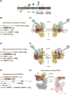
Figure 3.. Models of the Saccharomyces cerevisiae (A, B) and Schizosaccharomyces pombe (D) Dsc complex members.
Tul1 contains a N-terminal signal peptide (SP) sequence and a luminal domain with multiple glycosylation sites, seven predicted transmembrane domains and a C-terminal cytosolic RING domain. Rhomboid-like folds in Dsc2 are indicated with gray boxes. Dsc2 for S. pombe contains a C-terminal ubiquitin-associated (UBA) domain. Dsc3 contains an N-terminal ubiquitin-like (UBL). Ubx3 and Dsc5 contain an internal ubiquitin-associating (UAS) domain of unknown function and a C-terminal UBX domain that is known to interact with the AAA-ATPase Cdc48. Dsc5 contains an N-terminal coupling of ubiquitin to ER degradation (CUE) domain. Transmembrane domains (TMDs) are indicated with dark boxes and were predicted with DeepTMHMM. (A) Figure is adapted from Schmidt et al. [8]. (C, E) AlphaFold2 predictions of the indicated complexes and their members: Dsc1/Tul1 (gold), Dsc2 (red), Dsc3 (blue), Dsc4/Gld1 (brown), and Dsc5/Ubx3 (green). Resulting files were visualized with PyMOL, TMDs were predicted with DeepTMHMM and colored in dark. Left panel shows front view, right panel is rotated −120° around the y-axis. (F) Domains are indicated as in (B, D). Hrd3 contains three Haemolysin coregulated protein-like (HCP) domains. Crystal structure of Saccharomyces cerevisiae ERAD-L complexes. Left panel shows Der1 (red), Hrd1 (gold), and Hrd3 (gray) from PDB (6vjz). Right panel additionally shows Yos9 (pink) from PDB (6vk3). Files were visualized with PyMOL, TMDs were predicted with DeepTMHMM and colored in dark [81].
Figure 4.
Figure 4.. Rhomboid pseudo-proteases contain characteristic rhomboid-like folds.
(A–C) AlphaFold predictions of the indicated proteins were visualized with PyMOL. Transmembrane domains (TMDs) are colored in dark, amino-terminus (NH2), carboxy-terminus (COOH), and the loop region (L1) between TMD1 and TMD2 are indicated. (A) Dsc2 (AF-Q08232-F1). (B) Dfm1 (AF- Q12743-F1). (C) Der1 (AF-P38307-F1).

Similar articles

Cited by

  • Lysosomal quality control Review.
    Henn D, Yang X, Li M. Henn D, et al. Autophagy. 2025 Jul;21(7):1413-1432. doi: 10.1080/15548627.2025.2469206. Epub 2025 Feb 24. Autophagy. 2025. PMID: 39968899 Free PMC article. Review.

References

    1. Lemberg, M.K. and Strisovsky, K. (2021) Maintenance of organellar protein homeostasis by ER-associated degradation and related mechanisms. Mol. Cell 81, 2507–2519 10.1016/j.molcel.2021.05.004 - DOI - PubMed
    1. Christianson, J.C., Jarosch, E. and Sommer, T. (2023) Mechanisms of substrate processing during ER-associated protein degradation. Nat. Rev. Mol. Cell Biol. 24, 777–796 10.1038/s41580-023-00633-8 - DOI - PubMed
    1. Krshnan, L., van de Weijer, M.L. and Carvalho, P. (2022) Endoplasmic reticulum-associated protein degradation. Cold Spring Harb. Perspect. Biol. 14, a041247 10.1101/cshperspect.a041247 - DOI - PMC - PubMed
    1. Migliano, S.M., Wenzel, E.M. and Stenmark, H. (2022) Biophysical and molecular mechanisms of ESCRT functions, and their implications for disease. Curr. Opin. Cell Biol. 75, 102062 10.1016/j.ceb.2022.01.007 - DOI - PubMed
    1. Henne, W.M., Buchkovich, N.J. and Emr, S.D. (2011) The ESCRT pathway. Dev. Cell 21, 77–91 10.1016/j.devcel.2011.05.015 - DOI - PubMed

Publication types

Substances

LinkOut - more resources