. 2024 Nov 6;68(11):e0119424.
doi: 10.1128/aac.01194-24. Epub 2024 Sep 26.

Naamidine A reveals a promising zinc-binding strategy for topical antifungal therapy

Affiliations

Naamidine A reveals a promising zinc-binding strategy for topical antifungal therapy

Emily Puumala et al. Antimicrob Agents Chemother. .

Abstract

Fungal disease affects over a billion people worldwide. Naamidine A inhibits the growth of diverse fungal pathogens through an unknown mechanism. Here, we show that the supplementation of medium with excess zinc abolishes the antifungal activity of naamidine A. Furthermore, we highlight that naamidine A has in vitro activity against terbinafine-resistant Trichophyton spp. and in vivo efficacy in a mouse model of dermatomycosis caused by T. mentagrophytes, highlighting its therapeutic potential as a topical treatment.

Keywords: Candida albicans; Trichophyton; dermatomycosis; drug-resistant; metal chelation; natural product; zinc.


Conflict of interest statement

L.E.C. and L.W. are co-founders and shareholders in Bright Angel Therapeutics, a platform company for the development of novel antifungal therapeutics. L.E.C. is a Science Advisor for Kapoose Creek, a company that harnesses the therapeutic potential of fungi.

Figures
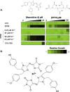
Fig 1
Fig 1
Zinc supplementation reduces the activity of naamidine A against C. albicans. (A) Twofold dose-response assays were performed against C. albicans (CaSS1) using naamidine A (0–25 µM), or diethylenetriamine penaacetate (DTPA; 0–100 µM) in 384-well plate format. The strain was grown overnight in YPD (1% yeast extract, 2% peptone, 2% dextrose) at 30°C, and then sub-cultured into YPD or RPMI medium (10.4 g/L RPMI powder with L-glutamine (Gibco), 165 mM MOPS, 2% glucose, 5 mg/mL histidine, pH 7) with metal supplementation or fetal bovine serum (FBS) as indicated. Plates were incubated at 30°C for 48 hours. Growth was monitored by optical density at 600 nm (OD600). The relative growth of treated samples was calculated by averaging technical duplicates and normalizing the OD600 of each treated well to the average OD600 of the corresponding untreated CaSS1 wild-type control. The results represent two independent biological replicates and were quantitatively plotted using Java TreeView3 (see color bar). (B) Proposed naamidine A-zinc coordination complex. Image generated using ChemDraw 19.0 (5, 6).
Fig 2
Fig 2
Naamidine A is active against drug-resistant T. indotiniae in culture and in a mouse model of T. mentagrophytes dermatomycosis. (A) Twofold dose-response assays were performed against T. indotiniae clinical isolates (CI) 633 /P/23 (terbinafine-sensitive), 18 (terbinafine-resistant), and V245-81 (terbinafine-resistant). Drug-susceptibility assays were performed in a 96-well format with twofold dilutions of naamidine A (0–50 µM) or terbinafine (0–110 µM) in RPMI with Zn+2 supplementation as indicated. T. indotiniae conidia were grown on PDA plates and harvested by washing with 10 mL saline-Tween solution (8 g/L NaCl, 0.025% Tween 20). Conidia were diluted to achieve a final assay cell density of 1 × 10⁶ CFU/mL and 200 ul was dispensed into wells. Plates were incubated in the dark at 28°C under static conditions for 96 hours. Relative fungal growth and viability were determined using alamarBlue reagent (Invitrogen; 1:20 dilution). Following the addition of alamarBlue, assay plates were incubated for 24 hours at 28°C. Fluorescence at 535/595 nm (Excitation/Emission) was read using a TECAN Spark multimode plate reader. The average relative fluorescence unit (RFU) value from media-only control wells was subtracted from all assay wells, and the data were normalized to the background-subtracted RFU in “no-compound” control wells. The plotted data are representative of two independent biological replicates and are quantitatively displayed as heat maps generated using Java TreeView3 (see color bar). (B) Male CD-1 mice were immunosuppressed with subcutaneous cortisone acetate (250 mg/kg) and intraperitoneal cyclophosphamide (200 mg/kg) at days −2, + 5, and +8 of infection. On the day of infection, mice were anesthetized with ketamine (82.5 mg/kg) and xylazine (6 mg/kg), their backs shaved, and a 3 × 3 cm area scraped gently with sandpaper. T. mentagrophytes suspension was then applied to denuded skin (17–19). At 5 days postinfection, twice daily treatment with 1% w/v naamidine A cream, control cream vehicle only, or no treatment was completed for 7 days. To quantify fungal burden in each group, mice were euthanized and resected infected skin samples were weighed, homogenized in PBS, and spread on PDA plates supplemented with 0.5 g/L chloramphenicol, 0.4 g/L cycloheximide, and 0.1% Triton X-100 (20). Plates were incubated at 30°C for 5 days. Fungal burden was quantified as CFU per gram of infected skin. The dashed line indicates the limit of detection. A Mann–Whitney test was used to determine statistical significance, and plots were generated using GraphPad Prism 9. Untreated: n = 8, Vehicle: n = 7, 1% Naamidine A: n = 9, ***P = 0.0002. (C) Representative photographs of skin sites infected with T. mentagrophytes at the completion of each treatment. All animal-related study procedures were compliant with the Animal Welfare Act, the Guide for the Care and Use of Laboratory Animals, and the Office of Laboratory Animal Welfare. The experiment was conducted under Protocol 31789 approved by the Lundquist Institute at Harbor-UCLA Medical Center.

References

    1. Bongomin F, Gago S, Oladele RO, Denning DW. 2017. Global and multi-national prevalence of fungal diseases-estimate precision. J Fungi (Basel) 3:57. doi:10.3390/jof3040057 - DOI - PMC - PubMed
    1. Fisher MC, Alastruey-Izquierdo A, Berman J, Bicanic T, Bignell EM, Bowyer P, Bromley M, Brüggemann R, Garber G, Cornely OA, Gurr SJ, Harrison TS, Kuijper E, Rhodes J, Sheppard DC, Warris A, White PL, Xu J, Zwaan B, Verweij PE. 2022. Tackling the emerging threat of antifungal resistance to human health. Nat Rev Microbiol 20:557–571. doi:10.1038/s41579-022-00720-1 - DOI - PMC - PubMed
    1. Puumala E, Fallah S, Robbins N, Cowen LE. 2024. Advancements and challenges in antifungal therapeutic development. Clin Microbiol Rev 37:e0014223. doi:10.1128/cmr.00142-23 - DOI - PMC - PubMed
    1. Martínez-Fructuoso L, Arends SJR, Freire VF, Evans JR, DeVries S, Peyser BD, Akee RK, Thornburg CC, Kumar R, Ensel S, Morgan GM, McConachie GD, Veeder N, Duncan LR, Grkovic T, O’Keefe BR. 2023. Screen for new antimicrobial natural products from the NCI program for natural product discovery prefractionated extract library. ACS Infect Dis 9:1245–1256. doi:10.1021/acsinfecdis.3c00067 - DOI - PMC - PubMed
    1. Vaden RM, Guillen KP, Salvant JM, Santiago CB, Gibbons JB, Pathi SS, Arunachalam S, Sigman MS, Looper RE, Welm BE. 2019. A cancer-selective zinc ionophore Inspired by the natural product naamidine A. ACS Chem Biol 14:106–117. doi:10.1021/acschembio.8b00977 - DOI - PMC - PubMed

Publication types

Supplementary concepts

LinkOut - more resources