. 2024 Oct 16;15(10):e0199124.
doi: 10.1128/mbio.01991-24. Epub 2024 Sep 26.

Increased intracellular H2S levels enhance iron uptake in Escherichia coli

Affiliations

Increased intracellular H2S levels enhance iron uptake in Escherichia coli

Shouta Nonoyama et al. mBio. .

Abstract

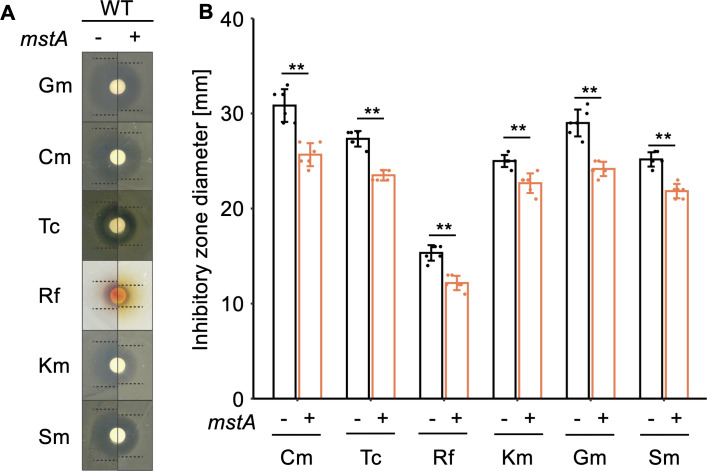
We investigated the impact of intracellular hydrogen sulfide (H2S) hyperaccumulation on the transcriptome of Escherichia coli. The wild-type (WT) strain overexpressing mstA, encoding 3-mercaptopyruvate sulfur transferase, produced significantly higher H2S levels than the control WT strain. The mstA-overexpressing strain exhibited increased resistance to antibiotics, supporting the prior hypothesis that intracellular H2S contributes to oxidative stress responses and antibiotic resistance. RNA-seq analysis revealed that over 1,000 genes were significantly upregulated or downregulated upon mstA overexpression. The upregulated genes encompassed those associated with iron uptake, including siderophore synthesis and iron import transporters. The mstA-overexpressing strain showed increased levels of intracellular iron content, indicating that H2S hyperaccumulation affects iron availability within cells. We found that the H2S-/supersulfide-responsive transcription factor YgaV is required for the upregulated expression of iron uptake genes in the mstA-overexpression conditions. These findings indicate that the expression of iron uptake genes is regulated by intracellular H2S, which is crucial for oxidative stress responses and antibiotic resistance in E. coli.

Importance: H2S is recognized as a second messenger in bacteria, playing a vital role in diverse intracellular and extracellular activities, including oxidative stress responses and antibiotic resistance. Both H2S and iron serve as essential signaling molecules for gut bacteria. However, the intricate intracellular coordination between them, governing bacterial physiology, remains poorly understood. This study unveils a close relationship between intracellular H2S accumulation and iron uptake activity, a relationship critical for antibiotic resistance. We present additional evidence expanding the role of intracellular H2S synthesis in bacterial physiology.

Keywords: Escherichia coli; YgaV; hydrogen sulfide; persulfide; reactive sulfur species; supersulfide.


Conflict of interest statement

The authors declare no conflict of interest.

Figures

Fig 1
Fig 1
Phenotype of the mstA-overexpressing strain. (A) Brown staining on Pb(Ac)2 soaked papers reflecting H2S synthesis in the WT strain harboring an empty vector (−) and/or the mstA-overexpression plasmid pMstA (+). (B) Quantification of brown-stained areas on Pb(Ac)2-soaked papers shown in panel A. The values are means ± SDs of three biological replicates. **P < 0.05, Welch’s t-test. (C) Volcano plot for upregulated and downregulated transcripts in the mstA-overexpression strain compared with those in WT harboring pUC18. Red dots represent transcripts within the significance threshold (P < 0.05, n = 3).
Fig 2
Fig 2
Antibiotic resistance of the mstA-overexpressing strain. (A) Photographs depicting the growth inhibitory circles around the antibiotic-soaked paper of the WT strain harboring an empty vector (−) and/or the mstA-overexpression plasmid pMstA (+). Dotted lines indicate the boundary of growth inhibitory circles. (B) Diameters of inhibitory circles shown in panel A, reflecting the antibiotic resistance of each strain. The values are means ± SDs of three biological replicates. **P < 0.05, Welch’s t-test.
Fig 3
Fig 3
Relationship between H2S overaccumulation-dependent and Fur-dependent transcript changes. Venn diagrams depict the overlap between genes upregulated (A) and downregulated (B) by mstA overexpression, and those regulated by apo- and holo-Fur. Genes directly regulated by apo- and holo-Fur, as reported previously (35), were used for the comparison, except for efeU_2 and ryhB, for which information was not found in the reference genome of the WT strain (BW25113) used in this study. (C) Intracellular iron content in WT harboring an empty vector (−) and/or the mstA-overexpression plasmid pMstA (+). The values are means ± SDs of five biological replicates. **P < 0.05, Welch’s t-test.
Fig 4
Fig 4
YgaV-dependent regulation of iron-uptake genes. Transcript levels of each gene were quantified by qRT-PCR. Total RNAs were extracted from the WT and/or ΔygaV mutant harboring an empty vector (−) and the mstA-overexpression plasmid pMstA (+). The values are means ± SDs of three biological replicates. Different letters (a, b, and c) indicate a significant difference (P < 0.05, Tukey’s test).
Fig 5
Fig 5
H2S-dependent YgaV binding to the target promoters as examined by ChIP-qPCR analysis. Binding of the His-tagged YgaV to the promoters of iron uptake genes (fes, fhuE, fhuF, and nfeF) in WT harboring an empty vector (−) and/or the mstA-overexpression plasmid pMstA (+) was examined as elaborated in Materials and Methods, with rrnB serving as a negative control gene. Pull-down of His-tagged YgaV for ChIP-qPCR was performed using a Ni-charged resin (Ni), as well as Ni-uncharged resin for a negative control (NC). The values are means ± SDs of three biological replicates. **P < 0.05, Welch’s t-test.
Fig 6
Fig 6
Iron import activity of the mstA-overexpressing strain. (A) Photographs depicting orange circles formed on CAS agar plates around the spotted bacterial cultures of the WT strain harboring an empty vector (−) and the mstA-overexpression plasmid pMstA (+). Dotted lines indicate the boundary of different colors. (B) Diameters of the orange circles shown in panel A, reflecting the iron transport activity of each strain. The values are means ± SDs of nine biological replicates. Different letters (a and b) indicate a significant difference (P < 0.05, Tukey’s test).
Fig 7
Fig 7
A model illustrating the H2S-mediated activation of iron uptake in E. coli.

Similar articles

Cited by

References

    1. Nishida M, Sawa T, Kitajima N, Ono K, Inoue H, Ihara H, Motohashi H, Yamamoto M, Suematsu M, Kurose H, van der Vliet A, Freeman BA, Shibata T, Uchida K, Kumagai Y, Akaike T. 2012. Hydrogen sulfide anion regulates redox signaling via electrophile sulfhydration. Nat Chem Biol 8:714–724. doi:10.1038/nchembio.1018 - DOI - PMC - PubMed
    1. Ono K, Akaike T, Sawa T, Kumagai Y, Wink DA, Tantillo DJ, Hobbs AJ, Nagy P, Xian M, Lin J, Fukuto JM. 2014. Redox chemistry and chemical biology of H2S, hydropersulfides, and derived species: implications of their possible biological activity and utility. Free Rad Biol Med 77:82–94. doi:10.1016/j.freeradbiomed.2014.09.007 - DOI - PMC - PubMed
    1. Shatalin K, Shatalina E, Mironov A, Nudler E. 2011. H2S: a universal defense against antibiotics in bacteria. Science 334:986–990. doi:10.1126/science.1209855 - DOI - PubMed
    1. Mironov A, Seregina T, Nagornykh M, Luhachack LG, Korolkova N, Lopes LE, Kotova V, Zavilgelsky G, Shakulov R, Shatalin K, Nudler E. 2017. Mechanism of H2S-mediated protection against oxidative stress in Escherichia coli. Proc Natl Acad Sci U S A 114:6022–6027. doi:10.1073/pnas.1703576114 - DOI - PMC - PubMed
    1. Ma Y, Yang X, Wang H, Qin Z, Yi C, Shi C, Luo M, Chen G, Yan J, Liu X, Liu Z. 2021. CBS-derived H2S facilitates host colonization of Vibrio cholerae by promoting the iron-dependent catalase activity of KatB. PLoS Pathog 17:e1009763. doi:10.1371/journal.ppat.1009763 - DOI - PMC - PubMed

MeSH terms

LinkOut - more resources