. 2024 Oct 16;15(10):e0156124.
doi: 10.1128/mbio.01561-24. Epub 2024 Sep 26.

Anaplasma phagocytophilum invasin AipA interacts with CD13 to elicit Src kinase signaling that promotes infection

Affiliations

Anaplasma phagocytophilum invasin AipA interacts with CD13 to elicit Src kinase signaling that promotes infection

Mary Clark H Lind et al. mBio. .

Abstract

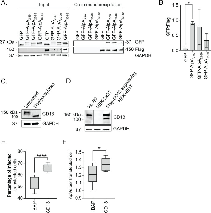
Host-microbe interactions that facilitate entry into mammalian cells are essential for obligate intracellular bacterial survival and pathogenesis. Anaplasma phagocytophilum is an obligate intracellular bacterium that invades neutrophils to cause granulocytic anaplasmosis. The invasin-receptor pairs and signaling events that induce Anaplasma uptake are inadequately defined. A. phagocytophilum invasion protein A orchestrates entry via residues 9-21 (AipA9-21) engaging an unknown receptor. Yeast two-hybrid screening suggested that AipA binds within C-terminal amino acids 851-967 of CD13 (aminopeptidase N), a multifunctional protein that, when crosslinked, initiates Src kinase and Syk signaling that culminates in endocytosis. Co-immunoprecipitation validated the interaction and confirmed that it requires the AipA N-terminus. CD13 ectopic expression on non-phagocytic cells increased susceptibility to A. phagocytophilum infection. Antibody blocking and enzymatic inhibition experiments found that the microbe exploits CD13 but not its ectopeptidase activity to infect myeloid cells. A. phagocytophilum induces Src and Syk phosphorylation during invasion. Inhibitor treatment established that Src is key for A. phagocytophilum infection, while Syk is dispensable and oriented the pathogen-invoked signaling pathway by showing that Src is activated before Syk. Disrupting the AipA-CD13 interaction with AipA9-21 or CD13781-967 antibody inhibited Src and Syk phosphorylation and also infection. CD13 crosslinking antibody that induces Src and Syk signaling restored infectivity of anti-AipA9-21-treated A. phagocytophilum. The bacterium poorly infected CD13 knockout mice, providing the first demonstration that CD13 is important for microbial infection in vivo. Overall, A. phagocytophilum AipA9-21 binds CD13 to induce Src signaling that mediates uptake into host cells, and CD13 is critical for infection in vivo.

Importance: Diverse microbes engage CD13 to infect host cells. Yet invasin-CD13 interactions, the signaling they invoke for pathogen entry, and the relevance of CD13 to infection in vivo are underexplored. Dissecting these concepts would advance fundamental understanding of a convergently evolved infection strategy and could have translational benefits. Anaplasma phagocytophilum infects neutrophils to cause granulocytic anaplasmosis, an emerging disease for which there is no vaccine and few therapeutic options. We found that A. phagocytophilum uses its surface protein and recently identified protective immunogen, AipA, to bind CD13 to elicit Src kinase signaling, which is critical for infection. We elucidated the AipA CD13 binding domain, which CD13 region AipA engages, and established that CD13 is key for A. phagocytophilum infection in vivo. Disrupting the AipA-CD13 interaction could be utilized to prevent granulocytic anaplasmosis and offers a model that could be applied to protect against multiple infectious diseases.

Keywords: Anaplasma; Anaplasmataceae; CD13; Rickettsiales; Src; Syk; bacterial internalization; host-pathogen interactions; invasin; obligate intracellular bacteria.


Conflict of interest statement

The authors declare no conflict of interest.

Figures

Fig 1
Fig 1
CD13 is an AipA interacting partner that benefits A. phagocytophilum infection. (A) AipA binds CD13. HeLa cells were transfected to express Flag-CD13 and GFP, GFP-AipA2–89, GFP-AipA22–89, or GFP-AipA32–89 and incubated with anti-Flag M2 affinity gel. Samples were analyzed by western blotting using antibodies specific for GFP, Flag, and glyceraldehyde-3-phosphate dehydrogenase (GAPDH). (B) The densitometric signals of co-immunoprecipitated GFP-tagged proteins were normalized to those of Flag. (C) Confirmation that CD13 is glycosylated in HL-60 cells. Uninfected HL-60 lysates were subjected to deglycosylation with PNGase F or not and analyzed by western blotting. (D) Expression of CD13 in HEK-293T cells. Whole-cell lysates from HL-60, HEK-293T, and Flag-CD13-expressing HEK-293T cells were lysed and analyzed by western blotting to assess the presence of CD13. (E and F) Ectopic expression of CD13 increases host cell permissiveness to A. phagocytophilum infection. HEK-293T cells expressing Flag-BAP or Flag-CD13 were incubated with A. phagocytophilum DC organisms. At 24 h, the cells were fixed, immunolabeled with A. phagocytophilum P44 and Flag antibodies, and examined by immunofluorescence microscopy to determine the percentage of infected transfected cells (E) and the number of ApVs per cell (F) in transfected cells. Results are representative of three independent experiments with similar results. Microscopy data are presented as box-and-whisker plots. The horizontal line denotes the median value (50th percentile). The gray boxes contain the 25th to 75th percentiles of the data set. The whiskers extend from the minimum to maximum values. Values beyond the upper and lower bounds are outliers indicated with black dots. One-way analysis of variance (ANOVA) with Tukey’s post hoc test was used to test for significant differences among groups. Student’s t test was used to test for a significant difference between pairs. Statistically significant values are indicated (*, P < 0.05; ****, P < 0.0001).
Fig 2
Fig 2
The CD13 C-terminus but not its enzymatic activity is important for A. phagocytophilum infection of HL-60 cells. (A–C) HL-60 cells were incubated with antibodies that bind PSGL-1 (KPL1), bind CD13781–967, bind the CD13 catalytic domain to inhibit its enzymatic activity (WM15), or isotype control. (D–F) HL-60 cells were treated with 1 mM bestatin or vehicle control (methanol). Treated HL-60 cells were incubated with A. phagocytophilum DC organisms. The cells were fixed, immunolabeled with P44 antibody, and analyzed at 1 h to determine the number of A. phagocytophilum (Ap) organisms bound per cell (A and D). The cells were examined at 24 h to determine the percentage of infected cells (B and E) and number of ApVs per cell (C and F). Results are indicative of three independent experiments. One-way ANOVA with Tukey’s post hoc test was used to test for significant differences among groups. Student’s t test was used to test for a significant difference between pairs. Statistically significant values are indicated (****, P < 0.0001).
Fig 3
Fig 3
The CD13 C-terminus but not its enzymatic activity is critical for A. phagocytophilum infection of human neutrophils. Human neutrophils were treated with the antibodies that bind PSGL-1 (KPL1), bind CD13781–967, bind the CD13 catalytic domain to inhibit its enzymatic activity (WM15), or isotype control and subsequently incubated with A. phagocytophilum DC organisms. The cells were fixed, immunolabeled with P44 antibody, and analyzed at 1 h to determine the number of A. phagocytophilum (Ap) organisms bound per neutrophil (A). The cells were examined at 24 h to determine the percentage of infected cells (B) and number of ApVs per cell (C). Results are indicative of three individual experiments. One-way ANOVA with Tukey’s post hoc test was used to test for a significant difference among groups. Statistically significant values are indicated (**, P < 0.01; ****, P < 0.0001).
Fig 4
Fig 4
Phosphorylation of Src kinase but not Syk is critical for A. phagocytophilum infection. (A) HL-60 cells were incubated with A. phagocytophilum DC bacteria after which samples were collected at 5 min, 30 min, 1 h, and 4 h. (B–D) HL-60 cells were treated with piceatannol (B), BAY (C), PP2 (D), or vehicle control dimethyl sulfoxide (DMSO) for 1 h and then incubated with DC organisms for 4 h in continued presence of the inhibitor. Samples were subjected to western blotting using antibodies against phospho-Syk (p-Syk; Y525/526) (A–D), Syk (A–D), phospho-Src (p-Src; Y416) (A–D), Src (A–D), phospho-Akt (p-Akt; S473) (B), Akt (B), A. phagocytophilum P44 (A–D), and GAPDH (A–D). Star denotes bands representative of phosphorylated Src. (E–G) HL-60 cells were treated with piceatannol, BAY, PP2, or DMSO and incubated with DC organisms. At 24 h post infection, the cells were fixed and immunolabeled with P44 antibody. Host cell nuclei and bacterial nucleoids were stained with DAPI. The samples were examined by immunofluorescence microscopy. (E) Representative merged fluorescent and brightfield micrographs are shown. The percentage of infected cells (F) and number of ApVs per cell (G) were determined. Results are representative of three independent experiments with similar results. One-way ANOVA with Tukey’s post hoc test was used to test for significant differences among groups. Statistically significant values are indicated (****, P < 0.0001).
Fig 5
Fig 5
CD13 crosslinking antibody fully restores infectivity of AipA antisera-treated A. phagocytophilum. (A) Confirmation that CD13 crosslinking mAb 452 increases levels of phospho-Src and phospo-Syk. HL-60 cells were incubated with isotype control or CD13 mAb 452 for 30 min. Samples were analyzed by western blotting using antibodies specific for phospho-Src (Y416), Src, phospho-Syk (Y525/526), Syk, and GAPDH. (B–D) mAb 452 rescues infectivity of AipA9–21 antisera-treated A. phagocytophilum. DC organisms treated with pre-immune sera or AipA9–21 antisera were incubated with HL-60 cells that had been pretreated with isotype control or mAb 452. Cells were analyzed by immunolabeling with A. phagocytophilum P44 to determine the number of A. phagocytophilum bound per cell at 1 h (B), the percentage of infected cells (C), and number of ApVs per cell (D) at 24 h. Results are representative of four independent experiments. Microscopy data are presented as box-and-whisker plots. One-way ANOVA with Tukey’s post hoc test was used to test for a significant difference among groups. Statistically significant values are indicated (****, P < 0.0001).
Fig 6
Fig 6
A. phagocytophilum engagement of CD13 elicits Src kinase and Syk phosphorylation. HL-60 cells were treated with isotype control or CD13781–967 antibodies and then incubated with A. phagocytophilum DC organisms. Uninfected HL-60 cells (U) were included as a control. Western blot and densitometric analyses were performed to detect phosphorylated and total levels of Src and Syk at the specified time points. (A) Western blots were probed for phospho-Src (Y416), Src, phospho-Syk (Y525/526), Syk, A. phagocytophilum P44, and GAPDH. Star indicates bands representative of phosphorylated Src. (B–D) The densitometric signals of phosphorylated Src, Syk, or P44 proteins normalized to that of GAPDH are presented. Data are representative of three independent experiments and presented as the mean ± SD. One-way ANOVA with Tukey’s post hoc test was used to test for significant differences among groups. Statistically significant values relative to isotype-treated cells are indicated (**, P < 0.01; ****, P < 0.0001).
Fig 7
Fig 7
The AipA9–21 receptor binding domain is critical for A. phagocytophilum to induce Src kinase and Syk phosphorylation. (A) Isolated A. phagocytophilum DC organisms were treated with AipA9–21 antibody or isotype control followed by incubation with HL-60 cells. Uninfected HL-60 cells (U) were included as a control. Western blot and densitometric analyses were performed to detect phosphorylated and total Src and Syk at the specified time points. Western blots were probed for phospho-Src (Y416), Src, phospho-Syk (Y525/526), Syk, A. phagocytophilum P44, and GAPDH. (B–D) The densitometric signals of phosphorylated Src, Syk, or P44 proteins were normalized to those of GAPDH. Data are representative of three independent experiments and presented as the mean ± SD. One-way ANOVA with Tukey’s post hoc test was used to test for significant differences among groups. Statistically significant values relative to isotype-treated cells are indicated (*, P < 0.05).
Fig 8
Fig 8
CD13 is required for A. phagocytophilum to productively infect mice. CD13 knockout (KO; seven females and five males) or wild-type (WT; five females and six males) mice were intraperitoneally injected with A. phagocytophilum DC organisms. Peripheral blood samples drawn on the indicated days were examined by light microscopy for A. phagocytophilum-infected neutrophils. Each symbol corresponds to the percentage of infected neutrophils as determined by examining at least 100 neutrophils per mouse. Two-way ANOVA with Tukey’s post hoc test was used to test for significant differences among groups. Data are presented as the mean ± SD. Statistically significant values are indicated (*, P < 0.05; **, P < 0.01; ****, P < 0.0001).
Fig 9
Fig 9
Working model of how the AipA-CD13 interaction contributes to A. phagocytophilum infection of host cells. The postulated mechanism by which AipA-CD13 and other A. phagocytophilum OMP-host cell receptor interactions mediate infection is based on findings presented herein and in references (4–12). (A) A. phagocytophilum engages sLex-capped PSGL-1 using OmpA and an unidentified OMP followed by AipA and Asp14 binding to CD13 and PDI, respectively. Together these interactions lead to phosphorylation of Src kinase followed by phospho-Src-dependent phosphorylation of Syk. Signaling through Src but not Syk promotes A. phagocytophilum infection, which can be inhibited by PP2 but not BAY. The AipA-CD13-induced Src phosphorylation and downstream events identified in this study are indicated by solid line arrows. Activation of Src via AipA- and CD13-independent interactions inferred from this study is indicated by dashed arrows. The A. phagocytophilum OMP that becomes thiol-reduced (star) by PDI and the host cell receptor that it subsequently binds are unknown. (B) AipA9–21 antibody blocks AipA binding to CD13 to prevent CD13-mediated Src signaling from promoting A. phagocytophilum uptake. Because other A. phagocytophilum-host cell receptor interactions also induce Src signaling, Src-dependent infection is compromised but not abolished. The addition of mAb 452, which crosslinks CD13 monomers to promote Src signaling in trans, restores the ability of anti-AipA9–21-treated A. phagocytophilum to infect. (C) CD13781–967 antibody binds the CD13 C-terminus to prevent A. phagocytophilum from crosslinking CD13 and thereby inhibit CD13-mediated Src signaling and bacterial uptake. Here again, CD13- and AipA-independent A. phagocytophilum-host cell interactions that also activate Src would facilitate infection, albeit at a reduced efficiency. (D) KPL1 antibody binds and prevents A. phagocytophilum interaction with the PSGL-1 N-terminus. The OmpA-sLex interaction alone is insufficient to dock the bacterium. KPL1 therefore robustly precludes AipA-CD13, Asp14-PDI, and any other host cell-pathogen interactions from forming to severely impair Src signaling and A. phagocytophilum uptake.

Similar articles

Cited by

References

    1. Atif FA. 2015. Anaplasma marginale and Anaplasma phagocytophilum: rickettsiales pathogens of veterinary and public health significance. Parasitol Res 114:3941–3957. doi:10.1007/s00436-015-4698-2 - DOI - PubMed
    1. MacQueen D, Centellas F. 2022. Human granulocytic anaplasmosis. Infect Dis Clin North Am 36:639–654. doi:10.1016/j.idc.2022.02.008 - DOI - PubMed
    1. Dixon DM, Branda JA, Clark SH, Dumler JS, Horowitz HW, Perdue SS, Pritt BS, Sexton DJ, Storch GA, Walker DH. 2021. Ehrlichiosis and anaplasmosis subcommittee report to the Tick-borne Disease Working Group. Ticks Tick Borne Dis 12:101823. doi:10.1016/j.ttbdis.2021.101823 - DOI - PubMed
    1. Troese MJ, Carlyon JA. 2009. Anaplasma phagocytophilum dense-cored organisms mediate cellular adherence through recognition of human P-selectin glycoprotein ligand 1. Infect Immun 77:4018–4027. doi:10.1128/IAI.00527-09 - DOI - PMC - PubMed
    1. Goodman JL, Nelson CM, Klein MB, Hayes SF, Weston BW. 1999. Leukocyte infection by the granulocytic ehrlichiosis agent is linked to expression of a selectin ligand. J Clin Invest 103:407–412. doi:10.1172/JCI4230 - DOI - PMC - PubMed

LinkOut - more resources