

Fazekas scale magnetic resonance imaging assessment in Alzheimer's disease and primary age-related tauopathy
- PMID: 39325192
- PMCID: PMC11611984
- DOI: 10.1007/s00234-024-03464-2
Fazekas scale magnetic resonance imaging assessment in Alzheimer's disease and primary age-related tauopathy
Abstract
Background: Brain vascular pathology is an important comorbidity in Alzheimer's disease (AD), with white matter damage independently predicting cognitive impairment. However, it is still unknown how vascular pathology differentially impacts primary age-related tauopathy (PART) compared to AD. Therefore, our objectives were to compare the brain microangiopathic burden in patients with PART and AD, evaluated by MRI, while assessing its relation with neuropathological findings, patterns of brain atrophy and degree of clinical impairment.
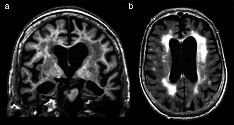
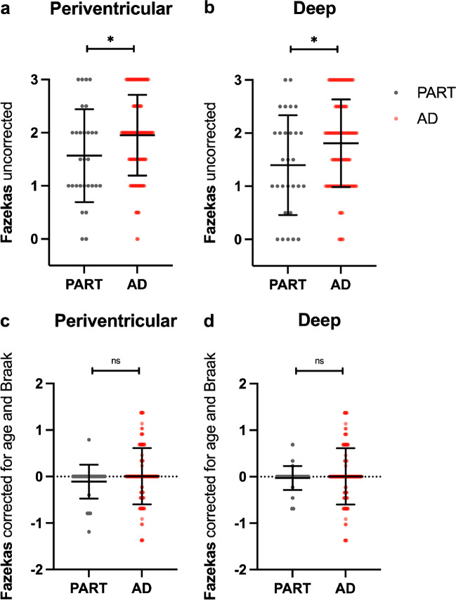
Methods: Clinical information, brain MRI (T1 and T2-FLAIR) and neuropathological data were obtained from the National Alzheimer's Coordinating Centre ongoing study, with a total sample of 167 patients identified, that were divided according to the presence of neuritic plaques in Consortium to Establish a Registry for Alzheimer's disease (CERAD) 0 to 3. Microangiopathic burden and brain atrophy were evaluated by two certified neuroradiologists, using, respectively, the Fazekas score and previously validated visual rating scales to assess brain regional atrophy.
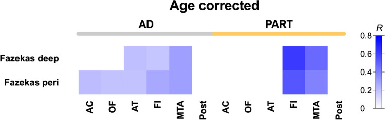
Results: Significant correlations were found between the Fazekas score and atrophy in the fronto-insular and medial temporal regions on both groups, with PART showing overall stronger positive correlations than in AD, especially in the fronto-insular region. For this specific cohort, no significant correlations were found between the Fazekas score and the degree of clinical impairment.
Conclusion: Our results show that PART presents different pathological consequences at the brain microvascular level compared with AD and further supports PART as an independent pathological entity from AD.
Keywords: AD = Alzheimer’s disease; CERAD = Consortium to Establish a Registry for Alzheimer’s disease; PART = Primary age-related tauopathy.
© 2024. The Author(s).
Conflict of interest statement
Declarations. Ethics approval: Study protocols were approved by the respective institutional review boards at each local Alzheimer’s Disease Centers. Informed consent: Written informed consent was provided by all participants at each local Alzheimer’s Disease Centers, and participants that contributed to the National Alzheimer’s Coordinating Center Neuropathology Data Set gave consent to autopsy. Competing interests: M.Q.N. is an Editorial Board Member of this journal, namely in Head and Neck/ENT Radiology Section. T.G.O. is a scientific advisor and shareholder of Ceracuity Inc, has been a consultant for Sonae and Guidepoint, has received fees as a speaker from Eisai and conference fees covered from Roche.
Figures
References
-
- Aa R (2019) Risk factors for Alzheimer’s disease. Folia Neuropathol 57(2):87–105. 10.5114/fn.2019.85929 - PubMed
MeSH terms
Grants and funding
LinkOut - more resources
Full Text Sources
Medical
