. 2024 Dec 2;134(23):e181612.
doi: 10.1172/JCI181612.

YTHDF1 loss in dendritic cells potentiates radiation-induced antitumor immunity via STING-dependent type I IFN production

Affiliations

YTHDF1 loss in dendritic cells potentiates radiation-induced antitumor immunity via STING-dependent type I IFN production

Chuangyu Wen et al. J Clin Invest. .

Abstract

The RNA N6-methyladenosine (m6A) reader YTHDF1 is implicated in cancer etiology and progression. We discovered that radiotherapy (RT) increased YTHDF1 expression in dendritic cells (DCs) of PBMCs from patients with cancer, but not in other immune cells tested. Elevated YTHDF1 expression in DCs was associated with poor outcomes for patients receiving RT. We found that loss of Ythdf1 in DCs enhanced the antitumor effects of ionizing radiation (IR) by increasing the cross-priming capacity of DCs across multiple murine cancer models. Mechanistically, IR upregulated YTHDF1 expression in DCs through stimulator of IFN genes/type I IFN (STING/IFN-I) signaling. YTHDF1 in turn triggered STING degradation by increasing lysosomal cathepsins, thereby reducing IFN-I production. We created a YTHDF1 deletion/inhibition prototype DC vaccine that significantly improved the therapeutic effect of RT and radioimmunotherapy in a murine melanoma model. Our findings reveal a layer of regulation between YTHDF1/m6A and STING in response to IR, which opens new paths for the development of YTHDF1-targeting therapies.

Keywords: Cancer immunotherapy; Dendritic cells; Immunology; Oncology; Radiation therapy.


Figures

Figure 1
Figure 1. Ythdf1 deficiency in DCs enhances the antitumor response to RT.
(A) MFI of YTHDF1 in DCs (CD11c+HLA-DR+) of PBMCs from patients with metastatic NSCLC with SBRT treatment via flow cytometry (n = 25). (B) Kaplan-Meier analysis of progression-free survival according to increased and unchanged YTHDF1 expression in DCs of PBMCs from NSCLC patients with SBRT treatment. (CF) WT (Ythdf1fl/fl) and Ythdf1-cKO (Cd11cCre Ythdf1fl/fl) mice were injected s.c. with MC38 (C and E) and B16-OZ (D and F) cells. Tumor-bearing mice were treated with local IR (1 dose) when the tumor volume reached 100–200 mm3. Tumor growth (C and D) and survival (E and F) were monitored after IR. Mice were considered dead if tumor volumes reached 2,000 mm3. Data are presented as the mean ± SEM. Data are representative of 2 or 3 independent experiments (CF). Two-sided, paired Student’s t test (A), 2-sided log-rank (Mantel-Cox) test (B, E and F), and 2-way ANOVA with Tukey’s multiple-comparison test (C and D). *P < 0.05, **P < 0.01, and ***P < 0.001.
Figure 2
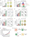
Figure 2. Ythdf1 deficiency increases the cross-priming capacity of DCs in the context of IR.
(A and B) WT and Ythdf1-cKO mice were injected s.c. with B16-OZ cells. Tumor-bearing mice were treated with local IR (20 Gy, 1 dose) when tumor volumes reached 100–200 mm3. On day 5 after IR, CD11c+ cells from TDLNs were isolated and cocultured with OT-I T cells for 3 days, and then IFN-γ–producing cells were enumerated by ELISPOT (n = 5) (A); in tumor-infiltrating DCs (CD45+F4/80CD11c+MHC-II+), the formation of H-2Kb-SIINFEKL was detected by flow cytometry (n = 5) (B). (CE). On day 8 after IR, the proportions of CD8+ T cells (CD45+CD3+CD8+) (C), IFN-γ (D), and granzyme B (E) in CD8+ T cells were detected by flow cytometry (n = 5). (F) On day 8 after IR, CD8+ T cells were isolated from TDLNs. Tumor antigen–specific CD8+ T cell function was measured via ELISPOT by coculturing CD8+ T cells with 5 μg/mL OT-I peptide (n = 5). (G) A dose of 200 μg anti-CD8 mAb was delivered twice weekly by i.p. injection to deplete CD8+ T cells, starting 1 day before IR. Tumor growth was monitored after IR. (H) Proposed model of how Ythdf1 KO in DCs sensitizes a tumor to IR by increasing the antitumor activity of CD8+ T cells. Data are presented as the mean ± SEM. Data are representative of 2 or 3 independent experiments. One-way ANOVA with Tukey’s multiple-comparison test (AF) and 2-way ANOVA with Tukey’s multiple-comparison test (G). *P < 0.05, **P < 0.01, and ***P < 0.001.
Figure 3
Figure 3. IR increases YTHDF1 expression in DCs via STING/IFN-I signaling.
(A) Violin plot showing the expression of Ythdf1 in total DCs from the scRNA-Seq data collected from tumor-infiltrating CD45+ cells of MC38-bearing mice on day 4 after IR. (BD). WT mice were injected s.c. with B16-OZ cells. Tumor-bearing mice received local IR (20 Gy, 1 dose) when the tumor volume reached 100–200 mm3. Tumor-infiltrating DCs (CD45+F4/80CD11c+MHC-II+) were sorted for detection of the expression of YTHDF1 on day 5 after IR via qPCR (n = 3) (B) and Western blotting (C). YTHDF1 expression in tumor-infiltrating DCs by intracellular staining and flow cytometry (n = 5) (D). (E and F) B16-OZ tumor–bearing Ifnar1-KO mice (E) and Sting-KO mice (F) received local IR (20 Gy, 1 dose), and the expression of YTHDF1 in tumor-infiltrating DCs was detected via flow cytometry on day 1 after IR (n = 4 or 5). (G and H) B16-OZ tumor–bearing Sting-KO mice (G) and Ifnar1-KO mice (H) were treated with 2 doses of 10 μg 2′3′-cGAMP (i.t.), and the expression of YTHDF1 of tumor-infiltrating DCs was detected by flow cytometry (n = 5). (I) Proposed mechanism of how IR induces YTHDF1 expression in DCs. IR-induced STING/IFN-I signaling increases the expression of YTHDF1 in DCs. Data are presented as the mean ± SEM. Data are representative of 2 or 3 independent experiments (BH) Wilcoxon Mann-Whitney U test (A), 2-sided, unpaired Student’s t test (B and D), and 1-way ANOVA with Tukey’s multiple-comparison test (EH). *P < 0.05, **P < 0.01, and ***P < 0.001.
Figure 4
Figure 4. Ythdf1 deletion enhances STING/IFN-I signaling in DCs.
(A) GSEA showing an increased IFN-β response in DCs with Ythdf1 deletion versus WT DCs. (B) WT and Ythdf1-cKO mice were injected s.c. with B16-OZ cells. Tumor-bearing mice underwent local IR (20 Gy, 1 dose) when the tumor volume reached 100–200 mm3, and tumors were excised on day 3 after IR for IFN-β ELISA (n = 5). (CE) BMDCs from Ythdf1-cKO mice were cocultured with 40 Gy–pretreated or nonirradiated B16-OZ cells for 8 hours. Purified CD11c+ cells were incubated for another 2 days. Supernatants were collected to measure IFN-β by ELISA (n = 3) (C). Purified CD11c+ cells were collected to measure mRNA levels of Ifnb (D) and Isg15, Ifit3, and Cxcl10 (E) by qPCR (n = 3). (F) BMDCs from Ythdf1-cKO and Ythdf1/Ifnar1-cKO mice were cocultured with B16-OZ cells for 8 hours. Purified CD11c+ cells were incubated with CD8+ T cells from OT-I mice for another 3 days. IFN-γ–producing cells were enumerated by ELISPOT (n = 3). (G) B16-OZ tumors in WT and Ythdf1-cKO mice were treated with IR and/or 200 μg anti-IFNAR1 mAb injected i.t. twice weekly. Tumor growth was monitored after IR. (H and I) BMDCs from Ythdf1-cKO and Ythdf1/Sting-cKO mice were cocultured with B16-OZ cells for 8 hours. Purified CD11c+ cells were incubated for an additional 2 days. IFN-β levels in supernatants were measured by ELISA (n = 3) (H). Purified CD11c+ cells were incubated with CD8+ T cells from OT-I mice for another 3 days. Then IFN-γ–producing cells were enumerated by ELISPOT (n = 3) (I). Data are presented as the mean ± SEM and are representative of 2 or 3 independent experiments. (BI) One-way ANOVA with Tukey’s multiple-comparison test (BF, H, and I) and 2-way ANOVA with Tukey’s multiple-comparison test (G). *P < 0.05, **P < 0.01, and ***P < 0.001.
Figure 5
Figure 5. Ythdf1 deletion diminishes the cathepsin-mediated decrease in STING expression in DCs.
(A) WT and Ythdf1-cKO mice were injected s.c. with B16-OZ cells. Tumor-bearing mice were treated with local IR (20 Gy, 1 dose) when the tumor volume reached 100–200 mm3. Expression of cathepsins A and B in tumor-infiltrating DCs (CD45+F4/80CD11c+MHC-II+) was detected via flow cytometry on day 5 after IR (n = 5). (B) BMDCs from WT mice were cocultured with 40 Gy–pretreated B16-OZ cells for 24 hours. Purified CD11c+ cells were collected to measure the enrichment of cathepsin A and B mRNA in the YTHDF1-immunoprecipitated RNA fraction (n = 3). (C) BMDCs from WT mice were treated with 2′3′-cGAMP and E64 for 12 hours, and STING expression was detected by Western blotting. (D) WT BMDCs and BMDCs with Ythdf1 deletion were treated with 2′3′-cGAMP for 12 hours, and STING expression was detected by Western blotting. (E) BMDCs from WT mice were stimulated with 2′3′-cGAMP. After immunoprecipitation with STING antibody, the expression of cathepsins A and B in whole-cell lysates was detected by Western blot. (F and G) BMDCs from WT mice were pretreated with E64 for 24 hours and then cocultured with 40 Gy–pretreated or nonirradiated B16-OZ cells for 8 hours. Purified CD11c+ cells were incubated for another 2 days. Supernatants were collected to measure IFN-β by ELISA (n = 3) (F), and cells were collected to measure Ifnb mRNA levels (n = 3) (G). (H) B16-OZ tumor–bearing WT mice were treated with local IR (20 Gy, 1 dose) and 50 μM E64 (i.t., daily). Tumor growth was monitored after IR. (I) Mechanism of how YTHDF1 affects STING/IFN-I signaling in DCs. IR-induced YTHDF1 increases the degradation of STING via cathepsins in the lysosomes of DCs, ultimately leading to decreased IFN-I production. Data are represented as the mean ± SEM and are representative of 2 or 3 independent experiments. Two-sided, unpaired Student’s t test (B), 1-way ANOVA with Tukey’s multiple-comparison test (A, F, and G), and 2-way ANOVA with Tukey’s multiple-comparison test (H). ***P < 0.001.
Figure 6
Figure 6. DC vaccines with YTHDF1 deletion/inhibition improve the response to RT and immunotherapy in murine cancers.
(A) Schematic diagram of the DC vaccine treatment plan. D, day. (B) WT mice were injected s.c. with B16-OVA cells. When the tumor volume reached 100–200 mm3, tumor-bearing mice were treated with IR (20 Gy, 1 dose) and antigen-pulsed BMDCs from WT or Ythdf1-KO mice (twice weekly by i.t. injection). (C) BMDCs from WT mice were treated with 20 μM SAC for 24 hours, and the expression of cathepsins A and B was detected by Western blotting. (D) BMDCs from WT mice were pretreated with SAC for 24 hours and then cocultured with 40 Gy–pretreated or nonirradiated B16-OZ cells for 8 hours. Purified CD11c+ cells were incubated for another 2 days, and the supernatants were collected to measure IFN-β (n = 3). (E) B16-OVA tumor–bearing mice were treated with IR (20 Gy, 1 dose) and antigen-pulsed BMDCs from WT mice with or without SAC treatment (twice weekly by i.t. injection). (F) B16-OVA tumor–bearing mice were treated with IR (20 Gy, 1 dose), antigen-pulsed BMDCs from Ythdf1-KO mice (twice weekly by i.t. injection), and anti–PD-L1 (twice weekly by i.p. injection). Data are represented as the mean ± SEM and are representative of 2 or 3 independent experiments. One-way ANOVA with Tukey’s multiple-comparison test (D) and 2-way ANOVA with Tukey’s multiple-comparison test (B, E, and F). *P < 0.05 and ***P < 0.001.

Comment in

  • Taking the STING out of radiotherapy: STING checkpoints mediate radiation resistance

References

    1. Zou Z, et al. The mechanism underlying redundant functions of the YTHDF proteins. Genome Biol. 2023;24(1):17. doi: 10.1186/s13059-023-02862-8. - DOI - PMC - PubMed
    1. Zaccara S, et al. Reading, writing and erasing mRNA methylation. Nat Rev Mol Cell Biol. 2019;20(10):608–624. doi: 10.1038/s41580-019-0168-5. - DOI - PubMed
    1. Shi H, et al. m6A facilitates hippocampus-dependent learning and memory through YTHDF1. Nature. 2018;563(7730):249–253. doi: 10.1038/s41586-018-0666-1. - DOI - PMC - PubMed
    1. Roundtree IA, et al. Dynamic RNA modifications in gene expression regulation. Cell. 2017;169(7):1187–1200. doi: 10.1016/j.cell.2017.05.045. - DOI - PMC - PubMed
    1. Wang X, et al. N(6)-methyladenosine modulates messenger RNA translation efficiency. Cell. 2015;161(6):1388–1399. doi: 10.1016/j.cell.2015.05.014. - DOI - PMC - PubMed

MeSH terms