. 2024 Sep 26;20(9):e1012576.
doi: 10.1371/journal.ppat.1012576. eCollection 2024 Sep.

The I7L protein of African swine fever virus is involved in viral pathogenicity by antagonizing the IFN-γ-triggered JAK-STAT signaling pathway through inhibiting the phosphorylation of STAT1

Affiliations

The I7L protein of African swine fever virus is involved in viral pathogenicity by antagonizing the IFN-γ-triggered JAK-STAT signaling pathway through inhibiting the phosphorylation of STAT1

Meilin Li et al. PLoS Pathog. .

Abstract

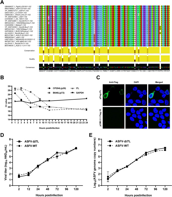
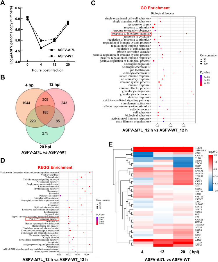
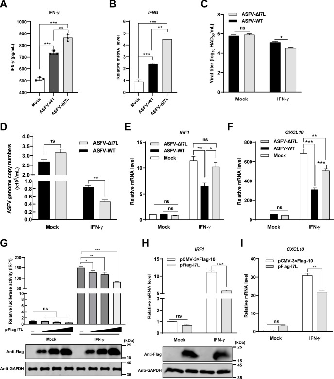
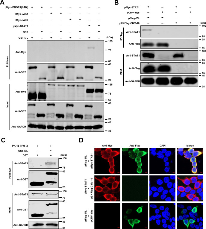
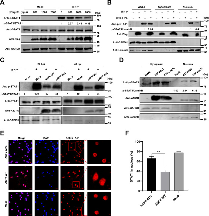
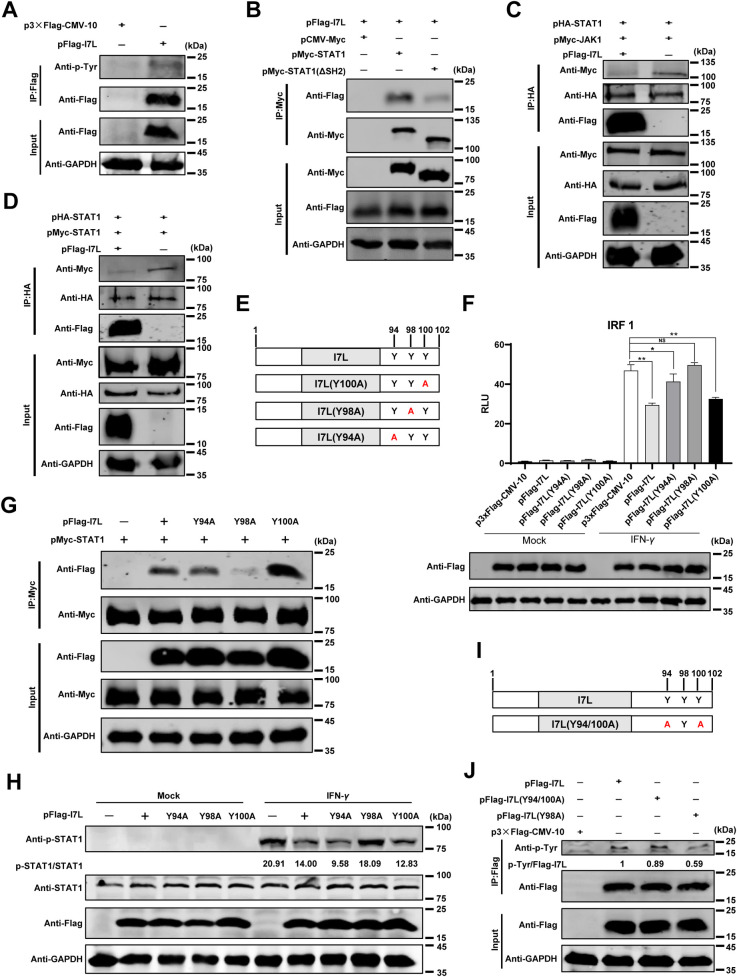
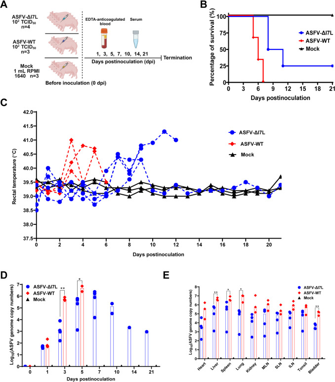
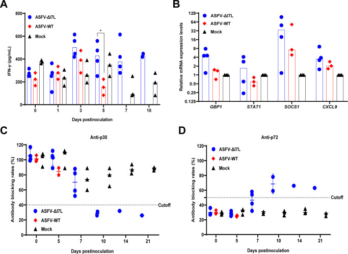
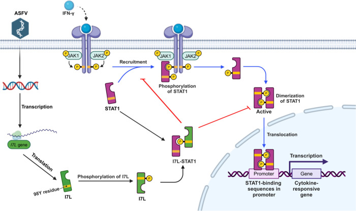
Cell-passage-adapted strains of African swine fever virus (ASFV) typically exhibit substantial genomic alterations and attenuated virulence in pigs. We have indicated that the human embryonic kidney (HEK293T) cells-adapted ASFV strain underwent genetic alterations and the I7L gene in the right variable region was deleted compared with the ASFV HLJ/2018 strain (ASFV-WT). A recent study has revealed that the deletion of the I7L-I11L genes results in attenuation of virulent ASFV in vivo, but the underlying mechanism remains largely unknown. Therefore, we hypothesized that the deletion of the I7L gene may be related to the pathogenicity of ASFV in pigs. We generated the I7L gene-deleted ASFV mutant (ASFV-ΔI7L) and found that the I7L gene deletion does not influence the replication of ASFV in primary porcine alveolar macrophages (PAMs). Using transcriptome sequencing analysis, we identified that the differentially expressed genes in the PAMs infected with ASFV-ΔI7L were mainly involved in antiviral immune responses induced by interferon gamma (IFN-γ) compared with those in the ASFV-WT-infected PAMs. Meanwhile, we further confirmed that the I7L protein (pI7L) suppressed the IFN-γ-triggered JAK-STAT signaling pathway. Mechanistically, pI7L interacts with STAT1 and inhibits its phosphorylation and homodimerization, which depends on the tyrosine at position 98 (Y98) of pI7L, thereby preventing the nuclear translocation of STAT1 and leading to the decreased production of IFN-γ-stimulated genes. Importantly, ASFV-ΔI7L exhibited reduced replication and virulence compared with ASFV-WT in pigs, likely due to the increased production of IFN-γ-stimulated genes, indicating that pI7L is involved in the virulence of ASFV. Taken together, our findings demonstrate that pI7L is associated with pathogenicity and antagonizes the IFN-γ-triggered JAK-STAT signaling pathway via inhibiting the phosphorylation and homodimerization of STAT1 depending on the Y98 residue of pI7L and the Src homology 2 domain of STAT1, which provides more information for understanding the immunoevasion strategies and designing the live attenuated vaccines against ASFV infection.


Conflict of interest statement

The authors have declared that no competing interests exist.

Figures

Fig 1
Fig 1. The biological characteristics of the I7L protein (pI7L).
(A) Multiple sequence alignment of pI7L among 20 ASFV strains. The homology analysis of pI7L was conducted using the Jalview software version 2.11.1.4. (B) The transcription dynamics of the I7L gene. Primary porcine alveolar macrophages (PAMs) were infected with the ASFV HLJ/2018 strain (ASFV-WT) at a multiplicity of infection of 1. At 1, 3, 6, 9, 12, and 24 hours postinfection (hpi), the average cycle threshold (CT) values of the PAMs were examined by a reverse transcription-quantitative PCR (RT-qPCR) using the primers targeting I7L, B646L (p72), CP204L (p30), and GAPDH. (C) Intracellular localization of pI7L. HEK293T cells were transfected with pFlag-I7L (0.5 μg). At 24 hpi, the cells were fixed and stained with a mouse anti-Flag monoclonal antibody and the nuclear marker DAPI and then examined by laser confocal microscopy. Bars, 5 μm. (D and E) The replication kinetics of ASFV-ΔI7L in PAMs. PAMs were seeded into 24-well plates and infected with ASFV-ΔI7L or ASFV-WT at a multiplicity of infection (MOI) of 0.01. At the indicated time points, the viral titers (D) and the genome copies (E) were determined by a quantitative real-time PCR (qPCR) and hemadsorption assay, respectively. Error bars denote the standard errors of the means. All the data were analyzed using the one-way ANOVA. ***, P < 0.001; ns, not significant.
Fig 2
Fig 2. Differential expression profiling in the ASFV-ΔI7L-infected primary porcine alveolar macrophages (PAMs) by RNA sequencing analysis.
(A) Identification of ASFV infection. PAMs were infected with ASFV-ΔI7L or ASFV-WT at a multiplicity of infection (MOI) of 1. The ASFV genome copies were determined by a quantitative real-time PCR (qPCR) at 4, 12, and 20 hours postinfection (hpi). (B) Venn diagrams of the differentially expressed genes (DEGs) in the ASFV-ΔI7L- versus (vs.) ASFV-WT-infected PAMs. (C and D) The bioinformatics analysis of DEGs. The gene ontology (GO) enrichment (C) and the Kyoto Encyclopedia of Genes and Genomes (KEGG) enrichment (D) analyses were performed in the ASFV-ΔI7L- vs. ASFV-WT-infected PAMs at 12 hpi. (E) Heat map of the DEGs induced by ASFV-ΔI7L vs. ASFV-WT at 4, 12, and 20 hpi. Error bars denote the standard errors of the means. All the data were analyzed using the one-way ANOVA. ***, P < 0.001; ns, not significant.
Fig 3
Fig 3. pI7L suppresses the IFN-γ-triggered JAK-STAT signaling pathway.
(A and B) ASFV-ΔI7L induces higher IFN-γ production than does ASFV-WT. Primary porcine alveolar macrophages (PAMs) were either mock-infected or infected with ASFV-ΔI7L or ASFV-WT at a multiplicity of infection (MOI) of 1. At 20 hours postinfection (hpi), the translational level of IFN-γ (A) and the transcriptional level of IFNG (B) were examined using the ELISA kit and reverse transcription-quantitative PCR (RT-qPCR), respectively. (C and D) pI7L antagonizes the antiviral activity of IFN-γ. PAMs were treated with IFN-γ or mock-treated for 24 hours, followed by infection with ASFV-ΔI7L or ASFV-WT at an MOI of 0.1 for 48 hours. The viral titers (C) and the genome copies (D) were determined by hemadsorption assay and quantitative real-time PCR (qPCR), respectively. (E and F) ASFV-ΔI7L induces higher IFN-γ-stimulated genes (ISGs) production than does ASFV-WT. PAMs were mock-infected or infected with ASFV-ΔI7L or ASFV-WT at an MOI of 1. At 24 hpi, the transcriptional levels of IRF1 (E) and CXCL10 (F) in the cell lysates were quantified by RT-qPCR. (G) pI7L inhibits the activation of the IRF1 promoter in a dose-dependent manner. HEK293T cells grown in 24-well plates were transfected with pFlag-I7L (0.1, 0.2, and 0.5 μg) together with the promoter reporter plasmid pIRF1-Fluc (100 ng) and the internal reference reporter plasmid pSV40-Rluc (10 ng). At 24 hours posttransfection (hpt), the transfected cells were mock-treated or treated with IFN-γ (20 ng/ml) for another 12 hours. The cells were lysed and the reporter activity was analyzed with a dual-luciferase assay kit. (H and I) pI7L inhibits the production of ISGs. HEK293T cells were transfected with pFlag-I7L or p3xFlag-CMV-10. At 24 hpt, the cells were mock-treated or treated with IFN-γ (20 ng/ml) for another 12 hours, and then the total RNA was extracted, and the transcriptional levels of IRF1 (H) and CXCL10 (I) in the cell lysates were quantified by a relative RT-qPCR. Error bars denote the standard errors of the means. All the data were analyzed using the one-way ANOVA. ***, P < 0.001; ns, not significant.
Fig 4
Fig 4. pI7L inhibits the IFN-γ-triggered JAK-STAT signaling pathway by targeting STAT1.
(A–C) pI7L interacts with STAT1. HEK293T cells were seeded into 6-well plates and transfected with pMyc-IFNGR1(ΔTM), -JAK1, -JAK2, or -STAT1. At 48 hours posttransfection (hpt), the cells were collected and lysed with NP-40 buffer. The purified GST or GST-pI7L protein was used to pull down the crucial adaptors of the JAK-STAT pathway and analyzed by Western blotting using a mouse anti-GST or -Myc monoclonal antibody (MAb) (A). HEK293T cells were cotransfected with pFlag-I7L and pMyc-STAT1. At 48 hpt, the lysates were collected and incubated with anti-Flag beads, and then the bound proteins were examined by Western blotting using a rabbit anti-Flag, -Myc, or -GAPDH MAb (B). PK-15 cells were treated with IFN-γ (100 ng/ml) for 12 hours and lysed with NP-40 buffer. The purified GST or GST-pI7L protein was used to pull down STAT1 and analyzed by Western blotting using a mouse anti-GST or -STAT1 MAb (C). (D) pI7L is colocalized with STAT1. For the confocal microscopy, the HEK293T cells were cotransfected with pFlag-I7L and pMyc-STAT1. At 24 hpt, the cells were incubated with a mouse anti-Flag or rabbit anti-Myc MAb and Alexa Fluor 488 (green)- or 594 (red)-conjugated secondary antibodies, respectively. The cell nuclei (blue) were stained with 4’,6-diamidino-2-phenylindole (DAPI). Bars, 5 μm.
Fig 5
Fig 5. pI7L inhibits STAT1 phosphorylation and blocks its nuclear translocation.
(A and B) pI7L inhibits the phosphorylation and the nuclear translocation of STAT1. HEK293T cells grown in 12-well plates were transfected with pFlag-I7L (500, 1000, or 2000 ng). At 24 hours posttransfection, the cells were treated with IFN-γ (100 ng/ml) or mock-treated for another 30 minutes, then the cells were lysed and the phosphorylation of STAT1 were examined by Western blotting using a rabbit anti-p-STAT1 (the phosphorylated STAT1), -STAT1, -Flag, or -GAPDH monoclonal antibody (MAb) (A). The pFlag-I7L–transfected cells were lysed and fractionated into cytoplasmic and nuclear fractions, and the p-STAT1 in the cytoplasmic and nuclear compartments were analyzed by Western blotting. Lamin B1 and GAPDH were used as nuclear and cytosolic markers, respectively. WCLs, whole cell lysates (B). (C–F) Deletion of the I7L gene promotes the phosphorylation and increases the nuclear translocation of STAT1. Primary porcine alveolar macrophages (PAMs) seeded into 12-well plates were infected with ASFV-ΔI7L or ASFV-WT at a multiplicity of infection (MOI) of 1 for 24 or 48 hours, followed by treatment with IFN-γ (100 ng/ml) for 30 minutes. The cells were lysed and the phosphorylation of STAT1 were analyzed by Western blotting using rabbit anti-p-STAT1, -STAT1, -A137R, or, -GAPDH MAbs (C). PAMs were mock-infected or infected with ASFV-ΔI7L or ASFV-WT at an MOI of 1 for 48 hours. The cells were lysed and fractionated into cytoplasmic and nuclear fractions, and the p-STAT1 in the cytoplasmic and nuclear compartments were analyzed by Western blotting. Lamin B1 and GAPDH were used as nuclear and cytosolic markers, respectively (D). PAMs were infected with ASFV-ΔI7L or ASFV-WT (MOI = 1) for 24 hours, and then treated with IFN-γ (100 ng/ml) for another 30 minutes. The subcellular localization of STAT1 (red) or cell nuclei (blue) was examined by laser confocal microscopy. Bars, 5 μm (E). The cells with STAT1 nuclear translocation were counted from 100 cells per condition from different view fields (F). Error bars denote the standard errors of the means. All the data were analyzed using the Student’s t test. **, P < 0.01.
Fig 6
Fig 6. pI7L inhibits the phosphorylation of STAT1 depending on its tyrosine at position 98.
(A) The phosphorylation modification occurs at the tyrosine residue of pI7L. HEK293T cells were transfected with pFlag-I7L or p3×Flag-CMV-10. At 24 hours posttransfection (hpt), the lysates were collected and incubated with anti-Flag beads, and then the bound proteins were examined by Western blotting using a rabbit anti-phosphotyrosine monoclonal antibody (MAb). (B) STAT1 interacts with pI7L depending on its SH2 domain. HEK293T cells were cotransfected pFlag-I7L with pMyc-STAT1 or pMyc-STAT1(ΔSH2). Co-IP assay was performed at 48 hpt using the anti-Flag beads. (C and D) pI7L inhibits the interaction between JAK1-STAT1 and STAT1-STAT1. HEK293T cells were cotransfected pFlag-I7L, pHA-STAT1 with pMyc-JAK1 (C) or pMcy-STAT1 (D). At 48 hpt, the lysates were collected and incubated with anti-HA beads, and then the bound proteins were checked by Western blotting using rabbit anti-Flag, -Myc, -HA, or -GAPDH MAb. (E) A schematic illustration of the tyrosine mutations in the C-terminus of pI7L. (F) pI7L(Y98A) fails to inhibit the activation of the IRF1 promoter. HEK293T cells grown in 24-well plates were transfected with p3xFlag-CMV-10, pFlag-I7L, -I7L(Y94A), -I7L(Y98A), or -I7L(Y100A) (0.5 μg each) together with the promoter reporter plasmid pIRF1-Fluc (100 ng) and the internal reference reporter plasmid pSV40-Rluc (10 ng), respectively. At 24 hpt, the transfected cells were treated with IFN-γ (20 ng/ml) or mock-treated for another 12 hours, and then the cells were lysed and the reporter activity was analyzed with a dual-luciferase assay kit. (G) pI7L interacts with STAT1 depending on the tyrosine at position 98. HEK293T cells were cotransfected p3xFlag-CMV-10, pFlag-I7L, -I7L(Y94A), -I7L(Y98A), or -I7L(Y100A) with pMcy-STAT1. At 48 hpt, the co-IP assay was performed. (H) pI7L(Y98A) does not inhibit the phosphorylation of STAT1. HEK293T cells grown in 12-well plates were transfected with p3xFlag-CMV-10, pFlag-I7L, -I7L(Y94A), -I7L(Y98A), or -I7L(Y100A). At 24 hpt, the cells were treated with IFN-γ (100 ng/ml) or mock-treated for another 30 minutes, then the cells were lysed and the phosphorylation of STAT1 was checked by Western blotting using rabbit anti-p-STAT1 (the phosphorylated STAT1), -STAT1, -Flag, or -GAPDH MAb. (I) A schematic illustration of the tyrosine mutations of pI7L(Y94/100A). (J) The phosphorylation occurs at the tyrosine residue at position 98 of pI7L. HEK293T cells were transfected with pFlag-I7L, pFlag-I7L(Y94/100A), or p3×Flag-CMV-10. At 24 hpt, the lysates were collected and incubated with anti-Flag beads, and then the bounded proteins were checked by Western blotting using a rabbit anti-phosphotyrosine MAb. All the data were analyzed using the one-way ANOVA. Error bars denote the standard errors of the means. *, P < 0.05; **, P < 0.01; ns, not significant.
Fig 7
Fig 7. The I7L gene is involved in ASFV pathogenicity in pigs.
(A) Schematic diagram of the pig inoculation experiment. The pigs were inoculated intramuscularly with ASFV-ΔI7L (n = 4, 102.0 HAD50/pig) or ASFV-WT (n = 3, 102.0 HAD50/pig) or mock inoculated with RPMI 1640 (n = 3, 1 mL/pig). The sera and the anticoagulated blood samples were collected at 0, 1, 3, 5, 7, 10, 14, and 21 days postinoculation. Created with BioRender.com. (B–E) pI7L is involved in ASFV pathogenicity in pigs. The survival rates (B) and rectal temperatures (C) of different groups were recorded. The viremia (D) and tissue viral loads (E) of each pig in different groups were quantified by a quantitative real-time PCR. MLN, mesenteric lymph node; SLN, submandibular lymph node; ILN, inguinal lymph node. All the data were analyzed using the Student’s t test. Error bars denote the standard errors of the means. *, P < 0.05; **, P < 0.01; ns, not significant.
Fig 8
Fig 8. ASFV-ΔI7L induces higher immune responses than does ASFV-WT in pigs.
(A and B) The ASFV-ΔI7L infection enhances the production of IFN-γ and IFN-γ-stimulated genes in pigs. The translational level of IFN-γ in the serum samples (A) and the mRNA transcriptional levels of GBP1, STAT1, SOCS1, and CXCL9 in the lung (B) were measured by ELISA kits and a reverse transcription-quantitative PCR, respectively. (C and D) The ASFV-ΔI7L infection induces serum antibodies in the inoculated pigs. The anti-p72 (C) or -p30 (D) antibodies in the ASFV-ΔI7L- or ASFV-WT-inoculated pigs were tested by ELISA kits. Error bars denote the standard errors of the means. All the data were analyzed using the Student’s t test. *, P < 0.05; ns, not significant.
Fig 9
Fig 9. A schematic model of the ASFV pI7L negatively regulating the JAK-STAT signaling pathway targeting STAT1 upon ASFV infection.
The ASFV pI7L can inhibit the IFN-γ-triggered JAK-STAT signaling pathway by targeting STAT1. pI7L interacts with STAT1 and inhibits the phosphorylation and homodimerization of STAT1 depending on the tyrosine at position 98 as well as the nuclear translocation of STAT1. Created with BioRender.com.

Similar articles

Cited by

References

    1. Galindo I, Alonso C. African swine fever virus: A review. Viruses. 2017; 9(5):103. doi: 10.3390/v9050103 - DOI - PMC - PubMed
    1. Sánchez-Cordón PJ, Montoya M, Reis AL, Dixon LK. African swine fever: A re-emerging viral disease threatening the global pig industry. Vet J. 2018; 233:41–48. doi: 10.1016/j.tvjl.2017.12.025 - DOI - PMC - PubMed
    1. Zhou X, Li N, Luo Y, Liu Y, Miao F, Chen T, et al.. Emergence of African swine fever in China, 2018. Transbound Emerg Dis. 2018; 65(6):1482–1484. doi: 10.1111/tbed.12989 - DOI - PubMed
    1. Gladue DP, Borca MV. Recombinant ASF live attenuated virus strains as experimental vaccine candidates. Viruses. 2022; 14(5):878. doi: 10.3390/v14050878 - DOI - PMC - PubMed
    1. Arabyan E, Kotsynyan A, Hakobyan A, Zakaryan H. Antiviral agents against African swine fever virus. Virus Res. 2019; 270:197669. doi: 10.1016/j.virusres.2019.197669 - DOI - PubMed

MeSH terms