Review
. 2024 Sep 27;385(6716):eadn6671.
doi: 10.1126/science.adn6671. Epub 2024 Sep 27.

Neuromodulator and neuropeptide sensors and probes for precise circuit interrogation in vivo

Affiliations
Review

Neuromodulator and neuropeptide sensors and probes for precise circuit interrogation in vivo

J Muir et al. Science. .

Abstract

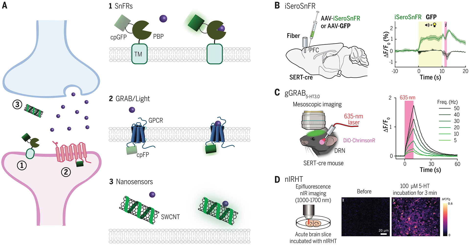
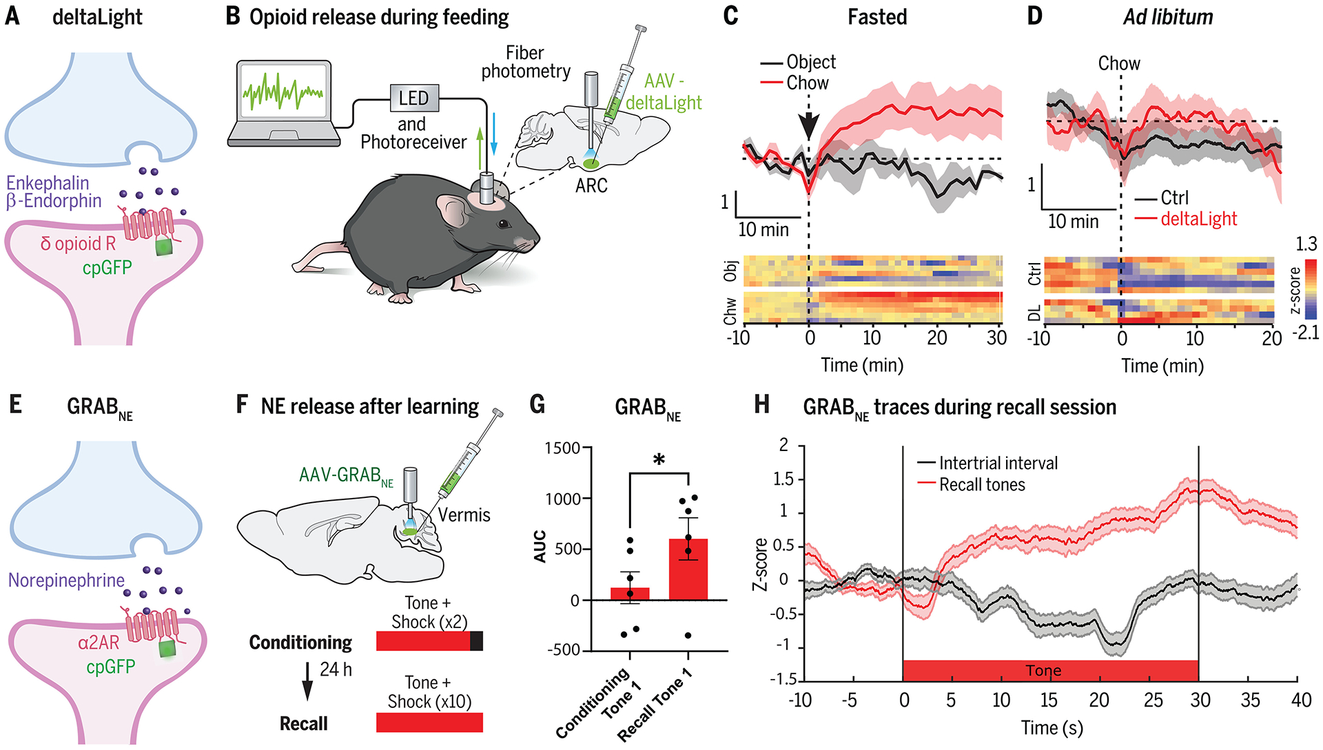
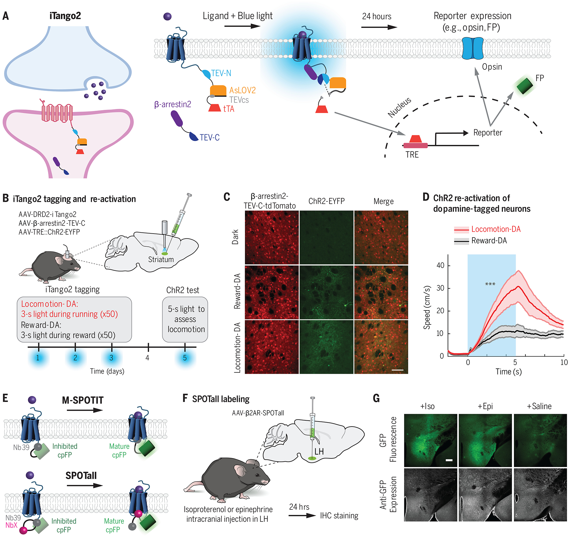
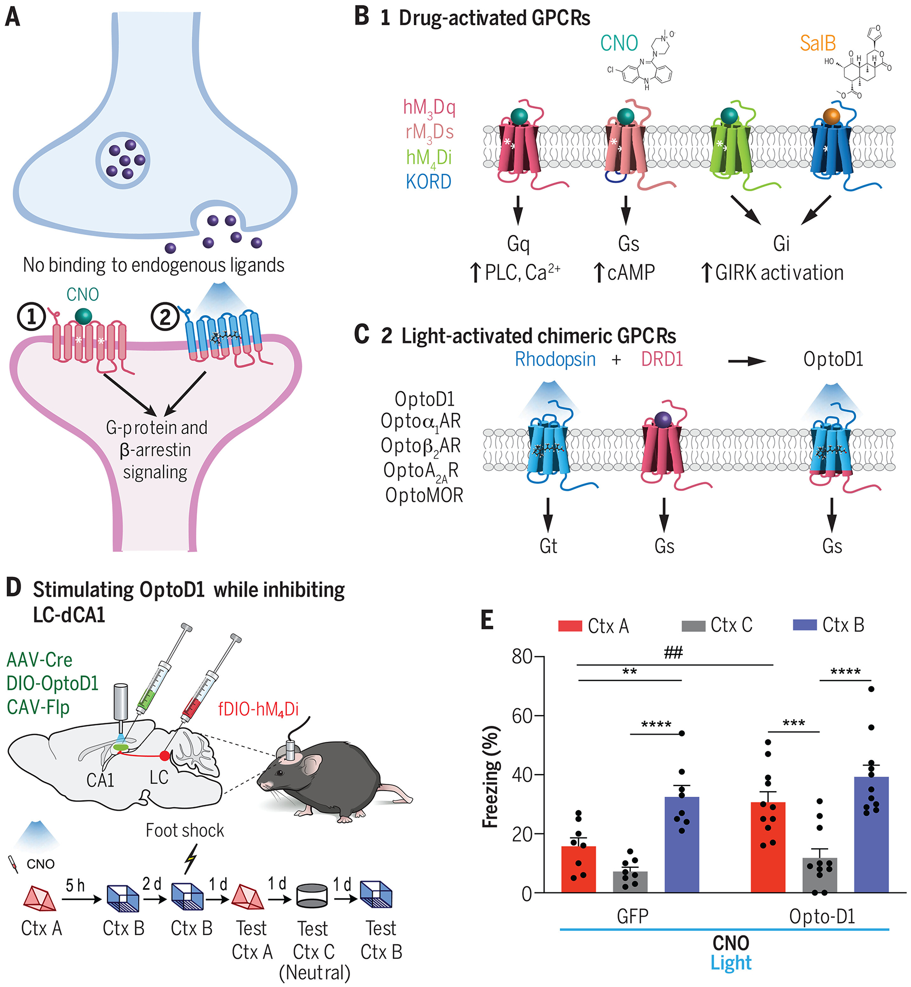
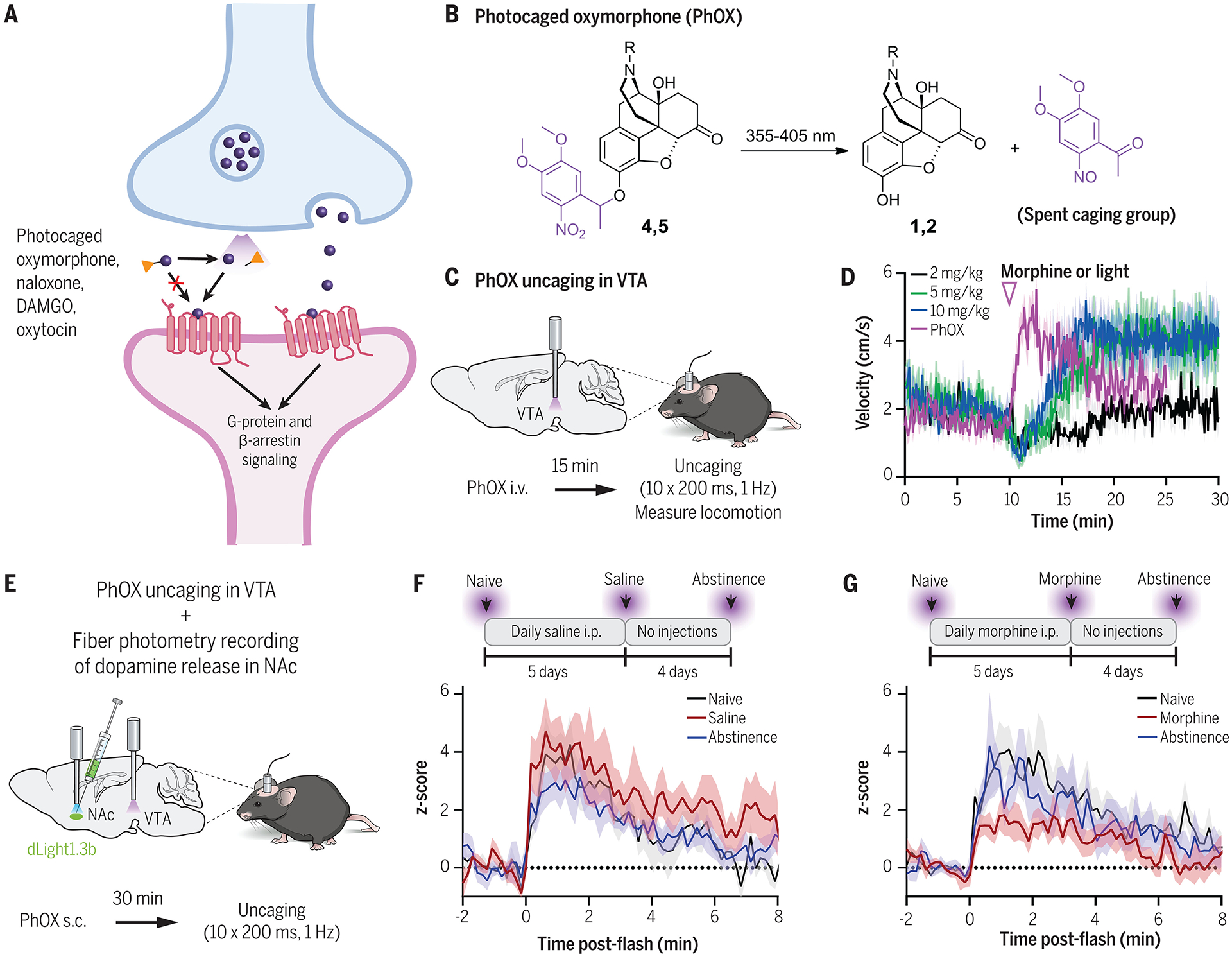
To determine how neuronal circuits encode and drive behavior, it is often necessary to measure and manipulate different aspects of neurochemical signaling in awake animals. Optogenetics and calcium sensors have paved the way for these types of studies, allowing for the perturbation and readout of spiking activity within genetically defined cell types. However, these methods lack the ability to further disentangle the roles of individual neuromodulator and neuropeptides on circuits and behavior. We review recent advances in chemical biology tools that enable precise spatiotemporal monitoring and control over individual neuroeffectors and their receptors in vivo. We also highlight discoveries enabled by such tools, revealing how these molecules signal across different timescales to drive learning, orchestrate behavioral changes, and modulate circuit activity.


Conflict of interest statement

Competing interests: Authors declare that they have no competing interests.

Figures

Fig. 1.
Fig. 1.. Fluorescent sensors for neuromodulator and neuropeptide release.
(A) Overview of main classes of sensor scaffolds used in brain tissue: 1. SnFRs are composed of a ligand-binding domain based on bacterial periplasmic binding proteins (PBP), which is fused to a circularly permutated GFP (cpGFP) and a transmembrane domain (TM). The cpGFP is located extracellularly. Upon ligand binding, a conformational change in the PBP binding pocket increases fluorescence emission of the cpGFP. 2. GRAB/Light sensors fuse a cpFP within the third intracellular loop of a GPCR. Ligand binding induces a conformational change leading to increased cpFP fluorescence. 3. Nanosensors are derived from polymer-functionalized semiconducting single-wall carbon nanotubes (SWCNTs), which are decorated with single stranded oligonucleotides that give the sensor affinity for specific ligands. Ligand binding alters the innate fluorescence of the SWCNT. (B) iSeroSnFR is a serotonin sensor based on an evolved PBP for acetylcholine. Because of its fast kinetics, iSeroSnFR can be used to record acute changes in serotonin dynamics during naturalistic behaviors such as aversive cue learning. Data shown from (57). (C) GRAB 5-HT3.0 (available as both a green and red version) is an improved serotonin sensor based on insertion of a cpFP into the third intracellular loop of the serotoning type 4 (5-HT4R) subtype. With a high affinity for serotonin, it can be used in vivo to monitor endogenous serotonin release. Either the red or green version can be combined with optogenetics to enable simultaneous read-out and control of serotonin signaling in vivo. Data shown from (75). (D) nIRHT is a SWCNT sensor for serotonin. It emits fluorescence in a nIR window at around 1200 nm. It has been validated in ex vivo mouse slice during bath serotonin application. Data shown from (86). See Table 1 for a list of genetically encoded neuromodulator and neuropeptide sensors designed for mammalian neuronal circuit studies.
Fig. 2.
Fig. 2.. Application of sensors to study neurochemical signaling in vivo.
(A) DeltaLight (67) is an engineered delta-opioid receptor that acts as a sensor for endogenous enkephalin and β-endorphin neuropeptides. (B) DeltaLight was targeted to the arcuate hypothalamic nucleus (ARC) by using an adeno-associated virus (AAV) injection to monitor opioid levels after feeding using fiber photometry. (C,D) DeltaLight fluorescence increases were observed in fasted mice after food presentation compared with presentation of an inedible object. However, non-fasted mice fed ad libitum did not show an increase in deltaLight fluorescence to food presentation. Data shown from (88). (E) GRABNE (78) is an engineered adrenergic receptor which detects endogenously released norepinephrine (NE). (F) GRABNE was injected into the cerebellar vermis of mice to record fluorescence emission during fiber photometry. Mice underwent a cued fear conditioning learning paradigm, and NE responses were recorded during the tone presentations. (G) There was an increase in NE release to the tone after fear conditioning, as measured by the area under the curve (AUC) of the GRABNE photometry traces. Data shown from (90). (H) Time course of NE release during the tone during the recall session (compared with intertrial intervals with no tone). Data shown from (90).
Fig. 3.
Fig. 3.. Molecular integrators for neuromodulator and neuropeptide release.
(A) iTango2 (95) is a transcriptional reporter system for detecting GPCR activation. In response to blue light and ligand-GPCR binding, a protease-mediated cleavage event occurs to release a membrane-bound transcription factor tTA. The tTA enters the nucleus where it drives expression of a TRE::reporter gene, such as an opsin or an FP. (B) DRD2-iTango2, β-arrestin2-TEV-C-TdTomato, and TRE::ChR2-EYFP viruses were injected bilaterally into the central striatum, and a fiberoptic cannula was implanted for blue light delivery. Water restricted mice were trained to run to receive a water reward. Blue light was delivered either during reward or locomotion across 3 days to tag neurons undergoing dopamine (DA) modulation. On a test day, blue light was delivered to activate ChR2-cells. ChR2, channelrhodopsin-2; EYFP, enhanced yellow fluorescent protein. (C,D) TRE::ChR2-EYFP expression was observed in mice tagged during either reward or locomotion. ChR2 stimulation of locomotion-DA neurons drove significantly more locomotion behavior compared with ChR2 stimulation of reward-DA neurons. Data shwon from (95). (E) M-SPOTIT and SPOTall (101) are integrators that rely on ligand-GPCR activation to drive the maturation of a fluorescent protein. cpGFP is fused to Nb39, a nanobody that binds the active μ-opioid receptor, and also happens to inhibit the maturation of cpGFP in this conformation. In M-SPOTIT, cpGFP-Nb39 is fused to the C terminus of the μ-opioid receptor. GPCR activation results in Nb39 binding, which no longer inhibits the cpGFP and thus allows it to mature and fluoresce. In SPOTall, different GPCR conformation-specific nanobodies (NbX) can be inserted between cpGFP and Nb39. Nb39 continues to block cpGFP maturation at rest, but upon ligand activation, NbX binds the GPCR and disrupts the Nb39-cpGFP interaction, resulting in enhanced GFP fluorescence. (F,G) SPOTall design for β2AR was tested in the lateral hypothalamus (LH) of mice. Elevated GFP fluorescence was observed in response to epinephrine (Epi) or isoproterenol (Iso) (which bind β2AR), but not in response to saline. Data shown from (101).
Fig. 4.
Fig. 4.. Examining behavioral effects using drug- and light-activated chimeric GPCRs.
(A) Overview of two classes of engineered GPCR actuators for inducing neuromodulator or neuropeptide receptor signaling in mammalian brain tissue. (B) General designs of commonly used designer receptors exclusively activated by designer drugs (DREADDs) (6, 113). DREADDs activate a specific downstream signaling cascade dependent on the engineered receptor. (C) Design strategy for creating light activated GPCRs (OptoXRs) (120). OptoXRs are engineered chimeras between a rhodopsin (which is activated by blue light) and the intracellular loops of the desired endogenously present GPCR. Upon blue light irradiation, the OptoXR undergoes conformational activation, triggering the native downstream intracellular signaling cascades specific to the desired GPCR. Depicted is OptoD1, which mimics DRD1 signaling in response to blue light (44). (D, E) Chowdhury et al. (129) simultaneously used an inhibitory DREADD and OptoXR to demonstrate the role of a dopaminergic locus coeruleus-to-dorsal hippocampal (LC-dCA1) circuit in memory linking. Whereas hM4Di inhibition of LC-dCA1 neurons disrupts fear memory linking across two contexts, simultaneous OptoD1 stimulation can rescue the memory linking deficits. CNO, clozapine-N-oxide; Ctx A, context A; Ctx B, context B, Ctx C, context C (neutral). Data shown from (129).
Fig. 5.
Fig. 5.. Photocaged ligands for spatiotemporal control of neurochemical signaling.
(A) Overview of photocaged ligands and peptides for acute neurochemical uncaging. In the absence of light, photoactivatable ligands are unable to bind to their receptors because of a photocleavable caging group. Light exposure cleaves off the caging group and allows the molecule to bind to endogenous receptors to trigger downstream signaling. (B) Chemical structure of a photocaged oxymorphone [PhOX (143)] before and after UV light activation. (C,D) PhOX uncaging in the VTA activates μ-opioid receptors and drives immediate behavioral changes in locomotion. Data shown from (143). (E-G) Simultaneous application of PhOX and a dopamine sensor, dLight1.3b (61), revealed how repeated morphine injections modulate opioid-induced VTA-to-NAc dopamine signaling in vivo. Dopamine release in the NAc was measured after acute PhOX activation in VTA at the naïve state (black), after 5 days of repeated saline (F) or morphine (G) injections (red), and after 4 days of abstinence from injections (blue). Mice undergoing repeated morphine injections showed a reduction in VTA opioid-NAc dopamine coupling. Data shown from (143).

Similar articles

Cited by

References

    1. Shen Y, Luchetti A, Fernandes G, Do Heo W, Silva AJ, The emergence of molecular systems neuroscience. Mol Brain 15, 7 (2022). - PMC - PubMed
    1. Zhang Y et al., Fast and sensitive GCaMP calcium indicators for imaging neural populations. Nature 615, 884–891 (2023). - PMC - PubMed
    1. Xu Y, Zou P, Cohen AE, Voltage imaging with genetically encoded indicators. Current Opinion in Chemical Biology 39, 1–10 (2017). - PMC - PubMed
    1. Deisseroth K, Optogenetics: 10 years of microbial opsins in neuroscience. Nature neuroscience 18, 1213 (2015). - PMC - PubMed
    1. Armbruster BN, Li X, Pausch MH, Herlitze S, Roth BL, Evolving the lock to fit the key to create a family of G protein-coupled receptors potently activated by an inert ligand. Proc Natl Acad Sci U S A 104, 5163–5168 (2007). - PMC - PubMed

Publication types