. 2025 Jan;246(1):120-133.
doi: 10.1111/joa.14142. Epub 2024 Sep 26.

Observations on the reproductive morphology of the female short-beaked echidna, Tachyglossus aculeatus

Affiliations

Observations on the reproductive morphology of the female short-beaked echidna, Tachyglossus aculeatus

Jane C Fenelon et al. J Anat. 2025 Jan.

Abstract

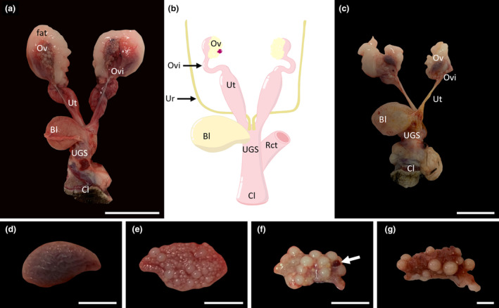
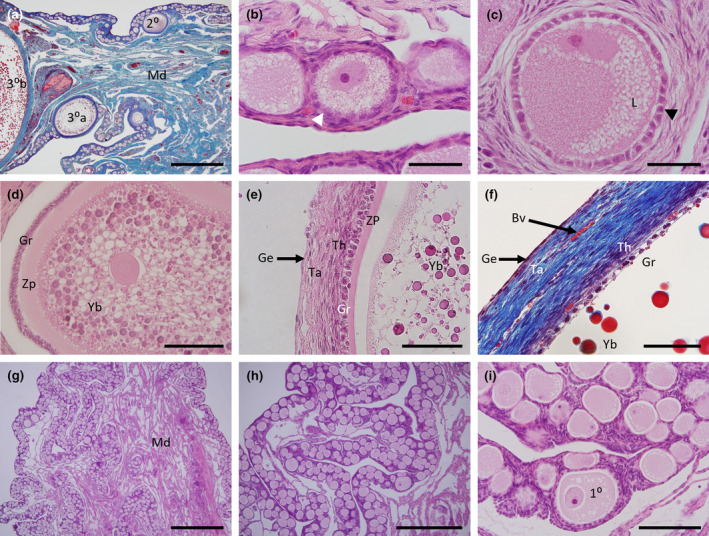
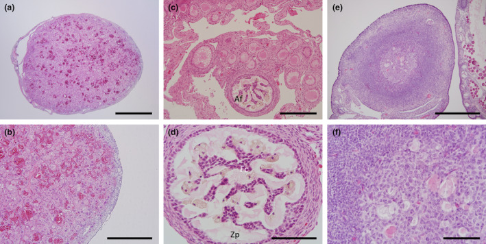
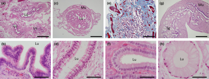
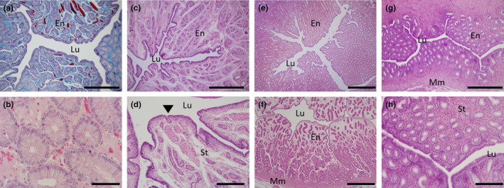
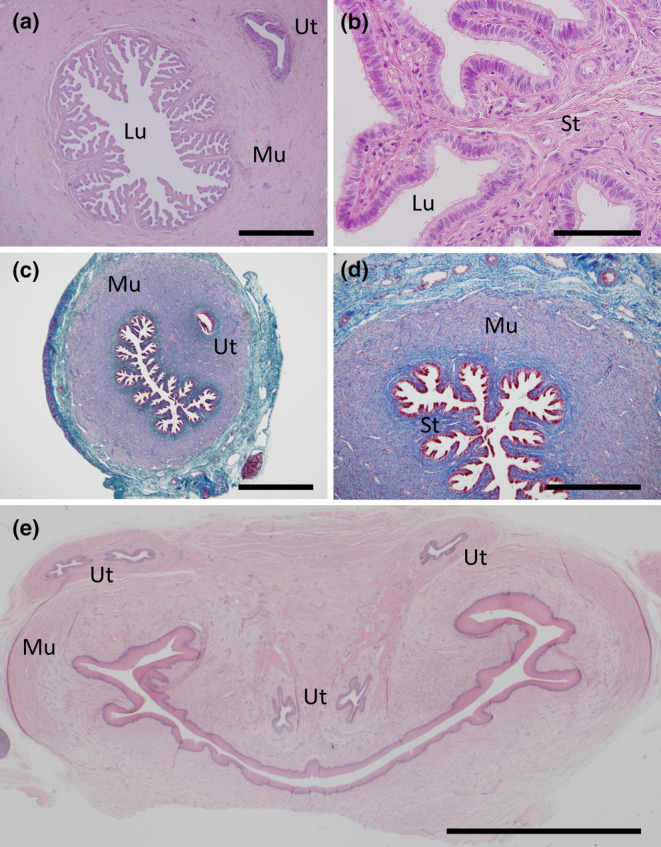
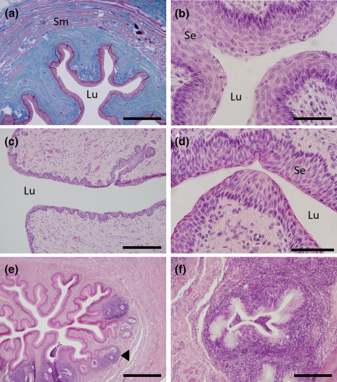
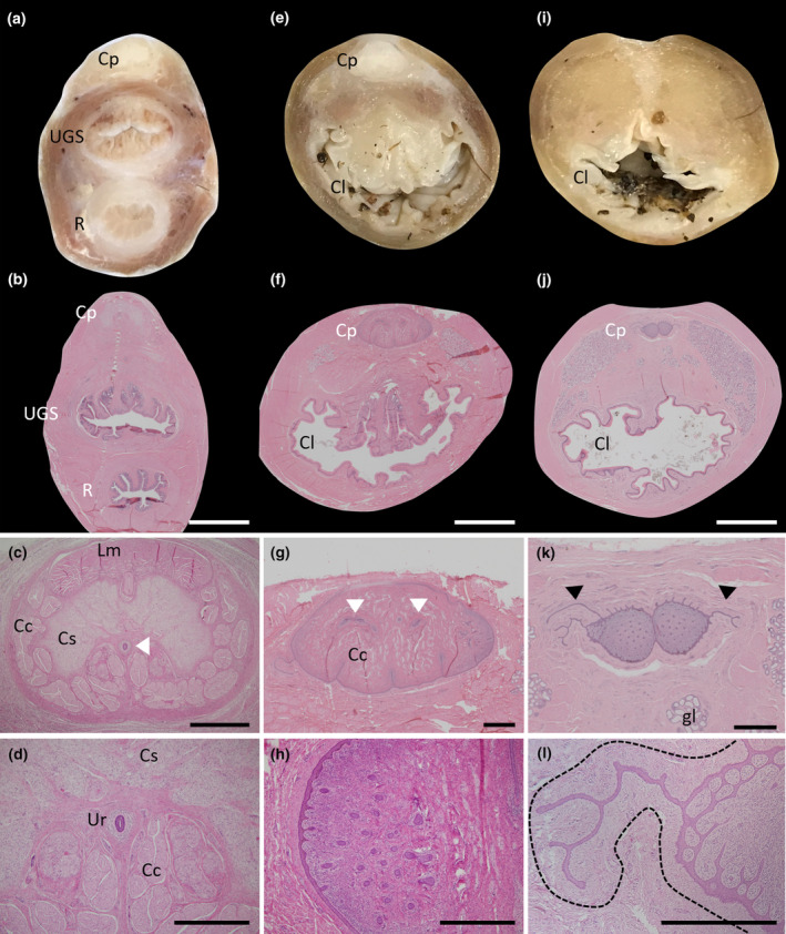
Although monotremes diverged from the therian mammal lineage approximately 187 million years ago, they retain various plesiomorphic and/or reptilian-like anatomical and physiological characteristics. This study examined the morphology of juvenile and adult female reproductive tracts across various stages of the presumptive oestrous cycle, collected opportunistically from cadaver specimens submitted to wildlife hospitals during the breeding season. In adult females, ovaries had a convoluted cortex with follicles protruding from the ovarian surface. While protruding antral follicles were absent from the ovaries of juvenile echidnas, histological analysis identified early developing primordial and primary follicles embedded into the ovarian cortex. The infundibulum epithelial cells of the oviducts were secretory during the follicular phase but not at other stages, the ampulla region was secretory at all stages and is likely responsible for the mucoid layer deposited around the zona pellucida, and the isthmus region of the oviduct appeared to be responsible for initial deposition of the shell coat, as in marsupials. Female echidnas have two separate uteri, which never merge and enter separately into the urogenital sinus (UGS). This study confirmed that both uteri are functional and increase in glandular activity during the luteal phase. In the juvenile uteri, the endometrium was immature with minimal, small uterine glands. A muscular cervical region at the caudal extremity of each uterus, just before the cranial region of the UGS was defined by the absence of glandular tissue in all female echidnas, including the juveniles. There was no evidence of a definitive vaginal region. A clitoris was also detected that possessed a less developed but similar structural (homologous) anatomy to the male penis; urethral ducts while present did not appear to be patent.

Keywords: cervix; monotreme; ovarian follicles; reproductive tract; uterine glands.


Figures

FIGURE 1
FIGURE 1
Overall morphology of the entire female echidna reproductive tract. (a) An adult female reproductive tract, showing well‐developed ovaries, oviducts, uteri, urogenital sinus (UGS) and cloaca. (b) Stylistic diagram of adult female reproductive tract highlighting the main reproductive structures and showing the position of the ureters and rectum relative to the tract. (c) Juvenile reproductive tract showing underdeveloped ovaries and narrow uterine tubes. (d) Morphology of an intact juvenile ovary, showing the smooth, convoluted structure of the ovary. (e) Morphology of an older juvenile showing multiple small and medium sized follicles. (f) Morphology of an intact adult ovary in the early luteal phase; white arrow identifies the location of a corpus luteum; (g): Morphology of an adult ovary in lactational anoestrus showing a reduced number of mature follicles and the convoluted cortex of the ovary. Bl, bladder; Cl, cloaca; Ov, ovary; Ovi, oviduct; Rct, rectum; UGS, urogenital sinus; Ur, ureter; Ut, uterus. Scale bars: (a) & (c) = 5 cm, (d–g) = 1 cm.
FIGURE 2
FIGURE 2
Histology of the echidna ovary and follicles. (a) Mallory's stained section of an adult female ovary showing the medulla (blue) with extensive blood vessels (red) and the narrow, convoluted ovarian cortex region containing multiple follicles at different stages of development. Note the presence of an early tertiary follicle (30a) and a partial section of a mature tertiary follicle (30b) on the far left. (b) H&E high power section of primordial follicles. (c) H&E high power section of a primary follicle with cuboidal granulosa cells and multiple lipid vacuoles within the follicle. (d) H&E section of a secondary follicle with numerous lipid bodies (white) and yolk bodies of varying sizes (dark pink). (e) H&E high power section of a mature tertiary follicle showing a layer of cuboidal granulosa cells and flattened thecal cells. (f) Mallory's stained high power section of a mature tertiary follicle showing the same structures as in (e) with the granulosa and thecal cell layers (purple) and surrounding connective tissue (blue) interspersed with red blood vessels and an external germinal epithelium (purple). (g) Low power H&E section of a juvenile ovary showing the medulla surrounded by the convoluted ovarian cortex. (h) H&E section of a juvenile ovary showing abundant follicles within the cortex. (i) High power image of (h) showing morphology of primordial and primary follicles. 10, Primary follicle; 20, Secondary follicle; 30a, Developing tertiary follicle; 30b, Mature tertiary follicle; Ge, Germinal epithelium; Gr, Granulosa cell layer; L, Lipid globules; Md, Medulla region; Ta, Tunica albuginea; Th, Thecal cell layer; Yb, Yolk bodies; Zp, Zona pellucida. Scale bars (a) & (g) = 1 mm, (b), (c) & (i) = 100 μm, (d) = 200 μm, (e) & (f) = 20 μm, (h) = 500 μm.
FIGURE 3
FIGURE 3
Histology of the corpora lutea and atretic follicles. (a) Section of a whole, mature corpora lutea. (b): High power image of (a) showing large round luteal cells packed densely with blood vessels. (c) Low power image showing size of an atretic early phase follicle. (d) High power image of (c) showing the pale pink staining convoluted fragmentation of the zona pellucida and invasion of the remnant follicular cells. (e) Atretic later phase follicle. (f) High power image of (e) showing remnants of the yolk bodies and granulosa cells. Af, atretic follicle; Fc, follicle cells; Zp, Zona pellucida. Scale bars: (a) & (c) = 1 mm, (b), (d) & (e) = 500 μm, (f) = 100 μm.
FIGURE 4
FIGURE 4
Histology of the adult oviduct. (a) H&E section of the three cross‐sections of the oviductal infundibulum region. (b) H&E high power image of the infundibulum showing the ciliated epithelium. (c) H&E transverse section of the ampulla region. (d) H&E high power image of the infundibulum epithelia from a recently ovulated female showing numerous secretory non‐ciliated goblet cells (pale staining) interspersed among the ciliated epithelial cells. (e) Mallory's stained sagittal section of the ampulla showing the high degree of vascularization (red) within the connective tissue (blue). (f) H&E high power image of the ampulla region with basal columnar, ciliated epithelium with no secretory vacuoles. (g) H&E section of the isthmus region with the appearance of the presumed shell glands; and thickened muscularis layer. (h) H&E high power image of a shell gland from the isthmus region. Bv, blood vessel; Lu, lumen; Mu, muscularis layer; Sg, shell glands. Scale bars: (a), (c), (e) & (f) = 1 mm, (b), (d), (f) & (h) = 50 μm.
FIGURE 5
FIGURE 5
Histology of the uterus. (a) Transverse section of a Mallory's stained uterus from an adult female showing the extent of the vascularization (red) in the stroma (blue) amidst the endometrial glands (purple); (b) H&E high power image of (a) showing the numerous enlarged uterine glands. (c) Transverse section of uterus from a lactating female with widely dispersed uterine glands. (d) High power image of (c) highlighting the small uterine glands relative to (b), arrowhead indicates a uterine gland exiting into the lumen. (e) Transverse section of uterus from a recently ovulated female with an active corpus luteum. (f) Transverse section of uterus from a female with a presumed inactive corpus luteum. (g) Transverse section of a juvenile echidna uterus. (h) High power image of (g) showing small uterine glands and densely packed stromal nuclei. En, endometrium; Lu, lumen; Mm, myometrium; St, endometrial stroma. Scale bars: (a), (c), & (g) = 500 μm, (e) & (f) = 1 mm, (b) = 20 μm and (d) & (h) = 200 μm.
FIGURE 6
FIGURE 6
Histology of the cervix. (a) H&E transverse section of an adult cervix showing the highly branched nature with no endometrial glands and the close proximity of the ureters. (b) H&E high power image of (a). (c) Mallory's stained transverse section of a juvenile cervix. (d) Mallory's high power image of (c) highlighting the minimal stroma present (dark blue) surrounded by an extensive muscularis (purple) and the surrounding connective tissue (light blue). (e) H&E transverse section of the upper urogenital sinus just after the merging of the left and right sides of the reproductive tract and the path of the left and right ureters just before their entry into the urogenital sinus. Lu, lumen; Mu, muscularis; St, stroma; Ut, ureter. Scale bars: (a), (c) = 1 mm, (b) = 100 μm, (d) = 500 μm, (e) = 5 mm.
FIGURE 7
FIGURE 7
Histology of the urogenital sinus (UGS). (a) Mallory's stained transverse section of the UGS showing the thickened stratified epithelium (red‐purple) surrounded by the sub‐mucosal connective tissue (blue) and the outer layers of smooth muscle (purple interspersed with blue). (b) H&E high power section of the UGS showing the thick layers of stratified squamous keratinized epithelium surrounding the urogenital sinus lumen. (c) H&E section of UGS from a female in lactational anoestrus showing the thinner layer of stratified squamous epithelium surrounding the lumen. (d) H&E higher magnification of the UGS epithelia of an early luteal phase female. (e) H&E transverse section of the UGS just above the rectum merging to form the cloaca. (f) H&E high magnification of the glandular structures in (e). Sm, smooth muscle; Se, stratified epithelium; Lu, lumen, black arrowhead indicates location of one of the glandular structures. Scale bars: (a) & (e) = 1 mm, (b) & (c) = 200 μm, (d) = 20 μm and (f) = 100 μm.
FIGURE 8
FIGURE 8
Morphology of the female clitoris. (a) Fixed section of the tract just above the merging of the rectum and UGS to form the cloaca showing the location of the clitoris. (b) H&E section of a similar area as (a). (c) Higher power section of the clitoral from (b). (d) High power image of the patent urethra. (e) Fixed section of the tract after the rectum and UGS have merged to form the cloaca. (f) H&E section of a similar area as (e). (g) Higher power section of the clitoris in (f) showing the branching of the urethra to each side (white arrowheads) and the radiation of the ducts on each edge. (h) High power image of the solid urethral duct radiations (dark purple). (i) Fixed section of the tract at the termination of the clitoris. (j) H&E section of a similar area to (i). (k) Higher power image of the clitoris in (j) showing the continual radiation of the solid urethral ducts (solid purple dots) and what appears to be the continued growth of the urethra into the tissue that immediately surrounds the clitoris (black arrowheads). (l) High power image of one of the urethral extensions into the tissue encasing the clitoris (defined by the black dotted line). Cp, clitoris (phallus‐like); Cl, cloaca; Cc, corpus cavernosum; Cs, corpus spongiosum; Gl, glandular tissue; Lm, levator muscle; R, rectum; UGS, urogenital sinus. Scale bars: (b), (f) & (j) = 5 mm, (c), (g) & (k) = 1 mm, (d), (h) & (l) = 500 μm.
FIGURE 9
FIGURE 9
Morphology of the female cloaca. (a) Fixed section of the cloaca just after the clitoris head has terminated. (b) H&E section of the cloaca in a similar area to (a) with extensive glandular tissue surrounding the cloacal lumen and the connection of the clitoral urethral extensions with the lumen of the cloaca (inset). (c) Higher power image of (b) showing the cloacal opening and the surrounding glandular tissue. (d) High power image of the glandular tissue. (e) Fixed section of the cloaca near to its terminal region with the pale coloured, discrete sebaceous glands visible. (f) Mallory's stained section of the cloaca in a similar area to (e) showing the keratinized epithelium (dark red), discrete sebaceous glands (pale purple) and glandular tissue below within the connective tissue (blue). (g) H&E higher power section of one of the sebaceous glands from (f) showing the central collection area of exudate and its connection with the lumen of the cloaca. (h) High power image of the sebaceous glandular tissue in (g). Ex, exudate; Gl, glandular tissue; Lu, lumen; Sg, sebaceous gland; Sm, smooth muscle. Scale bars: (b) & (f) = 5 mm, (c) & (g) = 1 mm and (d) & (h) = 200 μm.

Similar articles

References

    1. Allen NT (1982) A study of the hormonal control, chemical constituents, and functional significance of the paracloacal glands in Trichosurus vulpecula (including comparisons with other marsupials).). Perth, Australia: University of Western Australia.
    1. Augee, M.L. (2006) Echidna: extradordinary egg‐laying mammal. Collingwood, Victoria: CSIRO Publishing.
    1. Beard, L.A. , Grigg, G.C. & Augee, M.L. (1992) Reproduction by echidnas in a cold climate. In: Augee, M.L. (Ed.) Platypus and echidnas. Mosman, New South Wales, Australia: The Royal Zoological Society of NSW, Sydney, pp. 93–100.
    1. Buchtova, M. , Handrigan, G.R. , Tucker, A.S. , Lozanoff, S. , Town, L. , Fu, K. et al. (2008) Initiation and patterning of the snake dentition are dependent on Sonic hedgehog signaling. Developmental Biology, 319, 132–145. - PubMed
    1. Dutton‐Regester, K. , Keeley, T. , Fenelon, J.C. , Roser, A. , Meer, H. , Hill, A. et al. (2021) Plasma progesterone secretion during gestation of the captive short‐beaked echidna. Reproduction, 162, 267–275. - PubMed

Publication types

LinkOut - more resources