. 2024 Nov 13;44(46):e2184232024.
doi: 10.1523/JNEUROSCI.2184-23.2024.

Monosynaptic Inputs to Ventral Tegmental Area Glutamate and GABA Co-transmitting Neurons

Affiliations

Monosynaptic Inputs to Ventral Tegmental Area Glutamate and GABA Co-transmitting Neurons

Emily D Prévost et al. J Neurosci. .

Abstract

A unique population of ventral tegmental area (VTA) neurons co-transmits glutamate and GABA. However, the circuit inputs to VTA VGluT2+VGaT+ neurons are unknown, limiting our understanding of their functional capabilities. By coupling monosynaptic rabies tracing with intersectional genetic targeting in male and female mice, we found that VTA VGluT2+VGaT+ neurons received diverse brainwide inputs. The largest numbers of monosynaptic inputs to VTA VGluT2+VGaT+ neurons were from superior colliculus (SC), lateral hypothalamus (LH), midbrain reticular nucleus, and periaqueductal gray, whereas the densest inputs relative to brain region volume were from the dorsal raphe nucleus, lateral habenula, and VTA. Based on these and prior data, we hypothesized that LH and SC inputs were from glutamatergic neurons. Optical activation of glutamatergic LH neurons activated VTA VGluT2+VGaT+ neurons regardless of stimulation frequency and resulted in flee-like ambulatory behavior. In contrast, optical activation of glutamatergic SC neurons activated VTA VGluT2+VGaT+ neurons for a brief period of time at high frequency and resulted in head rotation and arrested ambulatory behavior (freezing). Stimulation of glutamatergic LH neurons, but not glutamatergic SC neurons, was associated with VTA VGluT2+VGaT+ footshock-induced activity and inhibition of LH glutamatergic neurons disrupted VTA VGluT2+VGaT+ tailshock-induced activity. We interpret these results such that inputs to VTA VGluT2+VGaT+ neurons may integrate diverse signals related to the detection and processing of motivationally salient outcomes.

Keywords: VTA VGluT2 neurons; co-transmit; colliculus; hypothalamus; threat; vesicular glutamate transporter 2.


Conflict of interest statement

The authors declare no competing financial interests.

Figures

Figure 1.
Figure 1.
Experimental design and VTA VGluT2+VGaT+ starter neurons. A, Schematic of viral injections for retrograde tracing in VGluT2::Cre/VGaT::Flp mice. B, Timeline of viral injections, tissue processing, and analysis. C–E, Example fluorescence images of viral injections in the VTA. C, VGluT2+VGaT+ neurons expressing the helper viruses. D, Neurons expressing the modified rabies virus. E, Neurons with mCherry-EGFP overlap (yellow triangle) coexpressed the helper viruses and rabies virus and comprised the population of starter neurons. Neurons with only mCherry expression (red triangle) did not receive the rabies virus and thus were not included in the starter neuron population. Neurons with EGFP only (green triangle) expressed the rabies virus and comprised the population of monosynaptic input neurons. F, Example histological section (top) registered to the Allen Mouse Brain Atlas using SHARCQ (bottom). G, Average proportion of starter neurons localized to the VTA and SuM. Dots represent individual animals; N = 8. H, Density heatmap of all starter neurons from −2.97 to −3.27 mm relative to the bregma (N = 8 animals). I, Coronal, sagittal, and horizontal 3D representations of all starter neurons (N = 8 animals). J, 3D coronal slice representations of starter neurons from (left to right) −2.37 to −2.67, −2.67 to −2.97, −2.97 to −3.27, −3.27 to −3.57, and −3.57 to −3.87 mm relative to the bregma (N = 8 animals). Abbreviations: SuM, supramammillary nucleus; VTA, ventral tegmental area.
Figure 2.
Figure 2.
Intersectional control experiment. A, Schematic of viral injections for retrograde tracing in VGluT2::Cre or VGaT::Flp mice. B, Example fluorescence image of viral injection in the VTA. C, Number of EGFP-expressing VTA neurons in VGluT2::Cre/VGaT::Flp, VGluT2::Cre, or VGaT::Flp mice. D, Number of EGFP and mCherry coexpressing VTA neurons in VGluT2::Cre/VGaT::Flp, VGluT2::Cre, or VGaT::Flp mice.
Figure 3.
Figure 3.
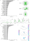
Brainwide monosynaptic inputs to VTA VGluT2+VGaT+ neurons. A, Average proportion of input neurons. All regions accounting for at least 0.5% of inputs are depicted. Dots represent individual animals; N = 8. B, Coronal, sagittal, and horizontal 3D representations of all input neurons (N = 8 animals). C, 3D coronal slice representations of input neurons from (left to right) 1.40 to 1.20, 0.20 to 0, −1.60 to −1.80, −3.15 to −3.35, −3.45 to −3.65, −4.30 to −4.50, and −5.8 to −6.0 mm relative to the bregma (N = 8 animals). D, Average density of input neurons. All regions with at least 10 neurons and a density of at least 100 neurons/mm3 are depicted. Dots represent individual animals; N = 8. E. Density heatmaps of input neurons in the DRN (top, −4.20 to −4.40 mm relative to the bregma), LHb (middle, −1.60 to −1.80 mm relative to the bregma), and PAG (bottom, −3.80 to −4.00 mm relative to the bregma). The bilateral LHb has a scale bar for each hemisphere. Abbreviations: DN, dentate nucleus; DRN, dorsal raphe nucleus; IP, interposed nucleus; LH, lateral hypothalamus; LHb, lateral habenula; MRN, midbrain reticular nucleus; NAcc, nucleus accumbens; PAG, periaqueductal gray; SC, superior colliculus; SNR, substantia nigra reticulata; VP/SI, ventral pallidum/substantia innominata; VTA, ventral tegmental nucleus.
Figure 4.
Figure 4.
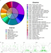
Cortical monosynaptic inputs to VTA VGluT2+VGaT+ neurons. A, Average proportion of input neurons in cortical regions. Interior pie chart illustrates broad cortical categories. Exterior pie chart illustrates subregions of cortical categories. B, 3D coronal slice representations of input neurons (N = 8 animals) from (left to right) 3.00 to 2.80, 2.50 to 2.30, 2.00 to 1.80, 1.50 to 1.30, 1.00 to 0.80, and 0.50 to 0.30 mm relative to the bregma. Abbreviations: ACA, anterior cingulate cortex; AI, agranular insular cortex; AON, anterior olfactory nucleus; M1, primary motor cortex; M2, secondary motor cortex; OFC, olfactory cortex; PIR, piriform area; S1, primary somatosensory cortex.
Figure 5.
Figure 5.
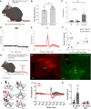
LH VGluT2+ neurons form a functional connection with VTA VGluT2+VGaT+ neurons and cause ambulatory behavior. A, Monosynaptic input neurons in LH. B, Bilateral density heatmap of input neurons in the LH from −1.60 to −1.80 mm relative to the bregma. The left scale bar corresponds to the left hemisphere; the right scale bar corresponds to the right hemisphere. C, D, Mice were first optically stimulated in an enclosed space. C, Schematic of viral injections and bilateral optical fiber implants for simultaneous optical stimulation of LH VGluT2+ neurons and fiber photometry of VTA VGluT2+VGaT+ neurons. D, Normalized averages (left) of GCaMP response to different frequencies of ChRmine laser stimulation (N = 4 animals). Average Z-scores (right) at stimulation were significantly higher than the baseline, invariant to stimulation frequency. Dots represent individual animals; N = 4. E–I, Mice were then optically stimulated in an unenclosed space while tail-restricted. E, Depiction of supported rearing and foot treading behavior elicited by ChRmine stimulation (wall not shown). F, Supported rearing and foot treading were significantly higher immediately following ChRmine stimulation compared with before stimulation. Dots represent individual animals; N = 4. G, Normalized average GCaMP response to 20 Hz ChRmine stimulation over 10 trials (N = 4 animals).
Figure 6.
Figure 6.
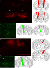
Histological localization of fiber optics targeting LH or SC VGluT2 neurons and VTA VGluT2+VGaT+ neurons. A, Example bilateral optical fiber placement and viral expression of AAV8-nEF-Con/Foff-ChRmine-oScarlet. B, Anatomical locations of all bilateral optical fiber cannulae (N = 4 animals). C, Example recording fiber placement and viral expression of AAV8-EF1-Con/Fon-GCaMP6m. D, Anatomical locations of all medial recording fiber cannulae (N = 4 animals). E, Example unilateral optical fiber placement and viral expression of AAV8-Nef-Con/Foff-ChRmine-oScarlet. F, Anatomical locations of all unilateral optical fiber cannulae (N = 4 animals). G, Example recording fiber placement and viral expression of AAV8-EF1-Con/Fon-GCaMP6m. H, Anatomical locations of all medial recording fiber cannulae (N = 4 animals). Scale bars in A and C, 100 µm.
Figure 7.
Figure 7.
SC VGluT2+ neurons form a functional connection with VTA VGluT2+VGaT+ neurons and reduce ambulatory behavior. A, Monosynaptic input neurons in the SC. B, Bilateral density heatmap of input neurons in the SC from −3.50 to −3.70 mm relative to the bregma. The left scale bar corresponds to the left hemisphere; the right scale bar corresponds to the right hemisphere. C, D, Mice were first optically stimulated in an enclosed space. C, Schematic of viral injections and unilateral optical fiber implant for simultaneous optical stimulation of SC VGluT2+ neurons and fiber photometry of VTA VGluT2+VGaT+ neurons. D, Normalized averages (left) of GCaMP response to different frequencies of ChRmine laser stimulation (N = 4 animals). Average Z-scores (right) depended on the interaction of frequency and stimulation condition. Dots represent individual animals; N = 4. E–J, Mice were then optically stimulated in an unenclosed space while tail-restricted. E, Depiction of head turning, freezing, and body turning behavior elicited by ChRmine stimulation. F, Normalized average GCaMP response to 40 Hz ChRmine stimulation over 10 trials (N = 4 animals). G, Proportion of stimulation trials and interstimulation intervals that resulted in head turning with freezing. Dots represent individual animals; N = 4. Head turning and freezing behavior was significantly more likely after stimulation compared with during interstimulation intervals. H, Average angle of head turn and body turn from starting position to freezing position following ChRmine stimulation for each test subject. Dots represent individual stimulation trials. All subjects except A518 had head turn angles normally distributed around their mean turn angle. Subjects A517 and A519 had body turn angles normally distributed around their mean turn angle, while A518 and A525 had body turn angle distributions that departed from normality. I, Box-and-whisker plot of head turn, peak GCaMP, and freeze latencies following ChRmine stimulation. Dots represent individual stimulation trials. Head turn latency was significantly shorter than peak GCaMP latency and freeze latency. Peak GCaMP and freeze latency did not differ. J, Average time spent freezing immediately following ChRmine stimulation and during interstimulation intervals, irrespective of pairing with head turning. The duration of continuous freezing bouts following stimulation (4.50 ± 0.33 s) was significantly longer than freezing in interstimulation intervals (0.33 ± 0.22 s). Dots represent individual animals; N = 4.
Figure 8.
Figure 8.
VTA VGluT2+VGaT+ neurons signal footshock outcomes that are correlated with LH VGluT2+ stimulation-induced VGluT2+VGaT+ activity. A, Schematic of viral injection and optical fiber implant for fiber photometry of VTA VGluT2+VGaT+ neurons from mice expressing ChRmine in either LH VGluT2 neurons or SC VGluT2 neurons. B, Wild-type mice show increased freezing behavior in response to a footshock-predicting CS+ compared with prior to CS+ presentation. Dots represent individual animals; N = 10. C, Normalized average maximum GCaMP response at the baseline, CS+, and footshock. Maximum GCaMP response was significantly higher at footshock compared with the baseline and CS+. Dots represent individual animals; N = 8. D, Normalized average GCaMP response at the CS+ onset (N = 8 animals). E, Normalized average GCaMP response at the footshock onset (N = 8 animals). F, VTA VGluT2+VGaT+ footshock-induced neuronal activity correlated with 40 Hz ChRmine stimulation-induced neuronal activity from LH VGluT2 neurons but not SC VGluT2 neurons. Dots represent individual animals; N = 8. G, Schematic for fiber photometry of VTA VGluT2+VGaT+ neurons in mice expressing inhibitory DREADD (hM4Di) or soma-tagged GtACR2 controls. Clozapine chemogenetically inhibits hM4Di but not stGtACR2 neurons. H, Example hM4Di expression in LH (left) and GCaMP expression in VTA (right). Scale bars, 200 µm. I, Cartoon of VTA optic fiber placements for hM4Di (red) and stGtACR2 (black) mice. J, Normalized average GCaMP response to clozapine and 0.3 mA tailshock (0.5 s duration). K, Tailshock increases GCaMP activity significantly above the baseline in control mice but not in hM4Di mice. Dots represent individual animals; N = 9.

Update of

References

    1. An S, et al. (2021) A whole-brain connectivity map of VTA and SNc glutamatergic and GABAergic neurons in mice. Front Neuroanat 15:818242. 10.3389/fnana.2021.818242 - DOI - PMC - PubMed
    1. Barbano MF, Wang HL, Zhang S, Miranda-Barrientos J, Estrin DJ, Figueroa-Gonzalez A, Liu B, Barker DJ, Morales M (2020) VTA glutamatergic neurons mediate innate defensive behaviors. Neuron 107:368–382.e8. 10.1016/j.neuron.2020.04.024 - DOI - PMC - PubMed
    1. Barbano MF, Zhang S, Chen E, Espinoza O, Mohammad U, Alvarez-Bagnarol Y, Liu B, Hahn S, Morales M (2024) Lateral hypothalamic glutamatergic inputs to VTA glutamatergic neurons mediate prioritization of innate defensive behavior over feeding. Nat Commun 15:403. 10.1038/s41467-023-44633-w - DOI - PMC - PubMed
    1. Basso MA, May PJ (2017) Circuits for action and cognition: a view from the superior colliculus. Annu Rev Vis Sci 3:197–226. 10.1146/annurev-vision-102016-061234 - DOI - PMC - PubMed
    1. Benarroch EE (2012) Periaqueductal gray: an interface for behavioral control. Neurology 78:210–217. 10.1212/WNL.0b013e31823fcdee - DOI - PubMed

MeSH terms

Substances

LinkOut - more resources