Review
. 2024 Sep 26;22(1):451.
doi: 10.1186/s12964-024-01779-4.

Immunomodulation in dengue: towards deciphering dengue severity markers

Affiliations
Review

Immunomodulation in dengue: towards deciphering dengue severity markers

Manoj Kumar Dash et al. Cell Commun Signal. .

Abstract

Background: Dengue is a vector-borne debilitating disease that is manifested as mild dengue fever, dengue with warning signs, and severe dengue. Dengue infection provokes a collective immune response; in particular, the innate immune response plays a key role in primary infection and adaptive immunity during secondary infection. In this review, we comprehensively walk through the various markers of immune response against dengue pathogenesis and outcome.

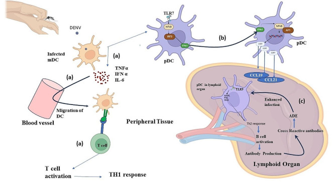
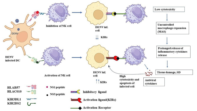
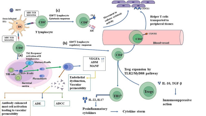
Main body: Innate immune response against dengue involves a collective response through the expression of proinflammatory cytokines, such as tumor necrosis factors (TNFs), interferons (IFNs), and interleukins (ILs), in addition to anti-inflammatory cytokines and toll-like receptors (TLRs) in modulating viral pathogenesis. Monocytes, dendritic cells (DCs), and mast cells are the primary innate immune cells initially infected by DENV. Such immune cells modulate the expression of various markers, which can influence disease severity by aiding virus entry and proinflammatory responses. Adaptive immune response is mainly aided by B and T lymphocytes, which stimulate the formation of germinal centers for plasmablast development and antibody production. Such antibodies are serotype-dependent and can aid in virus entry during secondary infection, mediated through a different serotype, such as in antibody-dependent enhancement (ADE), leading to DENV severity. The entire immunological repertoire is exhibited differently depending on the immune status of the individual.

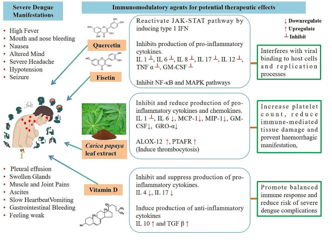
Short conclusion: Dengue fever through severe dengue proceeds along with the modulated expression of several immune markers. In particular, TLR2, TNF-α, IFN-I, IL-6, IL-8, IL-17 and IL-10, in addition to intermediate monocytes (CD14+CD16+) and Th17 (CD4+IL-17+) cells are highly expressed during severe dengue. Such markers could assist greatly in severity assessment, prompt diagnosis, and treatment.

Keywords: Aedes mosquitoes; Adaptive immunity; Dengue; Immunomodulator; Innate immunity.


Conflict of interest statement

The authors declare that they have no competing interests.

Figures

Fig. 1
Fig. 1
Innate immune response involving myeloid dendritic cells (mDCs). (a) Infected mDCs initiate the production of proinflammatory cytokines like TNF-α, IFN α, and IL-6 that are responsible for migration of mDCs to the site of infection (peripheral tissue), where the mDCs further activate the T lymphocytes and induce Th1 response. (b) Mitochondrial DNA (mtDNA) detected by TLR7 induces the expression of IFN-λ in other pDCs along with production of CCR-7. CCR-7 further acts as a receptor for CCL-19 and CCL-21 and assists in the migration of pDCs to lymphoid organs. (c) TLR9 detects mtDNA in pDCs that initiates signaling cascades involving MAPK p38 and NF-κB, thereby inducing Th2 response against DENV
Fig. 2
Fig. 2
Dengue virus non-structural protein 1(NS1) inhibits natural killer (NK) cell cytotoxic effects. Dengue NS1 restricts the KIR3DL1 receptor on NK cell and prevents KIR-MHC binding, which further inhibits NK cell function resulting in reduced cytotoxicity. Reduced cytotoxicity causes macrophage hyperactivation and proinflammatory cytokine production leading to tissue damage and clinical severity. On the contrary, NS3 binds with KIR3DL2 receptor and leads to the activation of NK cells, leading to high cytotoxicity and rapid clearance of DENV-infected cells
Fig. 3
Fig. 3
Adaptive T-cell response to dengue virus (DENV) infection. (a) Dendritic cell (DC) presentation of the DENV antigen activates CD4+T cells that develop into T follicular helper (TfH) cells. TfH cell further migrates to the periphery of the B cell follicle in the lymph nodes, where they take part in the germinal center reaction that fosters the growth of DENV-specific plasma cells and memory B cells. These plasma cells enhance the production of antibodies that aid in mast cell activation and activate antibody-dependent cellular cytotoxicity (ADCC) and antibody dependent enhancement (ADE) during secondary DENV infection. (b) Activated CD4+T cells also lead to the development of T regulatory (Tregs) and T helper 17 (Th17) cells. Th17 cells are then transported to peripheral tissues. (c) CD8 + T cells after activation move directly to the peripheral tissues leading to cytotoxic activity
Fig. 4
Fig. 4
Mechanistic effects of complex interplay between pro-inflammatory and anti-inflammatory cytokines and chemokines during dengue virus (DENV) infection. (a) Infection of DENV to host via Aedes mosquito during blood meal. (b) DENV infects target cells including dendritic cells, macrophages, and monocytes, initiating host immune response. (c) Infected cells release pro-inflammatory cytokines, such as IL-1, IL-6, IL-8, IL-17, IL-12, TNF-α, and GM-CSF and chemokines such as MCP-1/CCL-2, MIP-1β/CCL-4, CXCL-8, and GRO-α/CXCL-1. Proinflammatory cytokines activate other immune cells and promote the release of anti-inflammatory cytokines, such as IL-10 and TGF-β that limits the extent of immune responses. During high dengue viremia, excessive release of pro-inflammatory cytokines (IL-1, IL-6, IL-8, IL-17, IL-12, TNF-α, and GM-CSF) and chemokines (MCP-1/CCL-2, MIP-1β/CCL-4, CXCL-8, and GRO-α/CXCL-1), upregulation of anti-inflammatory cytokines (IL-10 and TGF-β), and downregulation of chemokine RANTES/CCL-5 lead to exaggerated immune response. This surge in immune response results in cytokine storm and contribute to SD manifestations
Fig. 5
Fig. 5
Mechanistic figure showing potential therapeutic effects of antioxidants including quercetin, fisetin, Carica papaya leaf extract, and immunomodulatory agent vitamin D on the cytokine and chemokine responses in suppressing severe dengue manifestations

References

    1. Heilman JM, De Wolff J, Beards GM, Basden BJ, Heilman J. Dengue fever: a Wikipedia clinical review. Open Med. 2014. - PMC - PubMed
    1. Fong S-L, Wong K-T, Tan C-T. Dengue virus infection and neurological manifestations: an update. Brain. 2024;147:830–38. - PubMed
    1. Dengue and severe dengue [cited 2024 Apr 25]. Available from: https://www.who.int/news-room/fact-sheets/detail/dengue-and-severe-dengue
    1. Kumar M, Verma R, Nirjhar S, Singh M. Dengue in children and young adults, a cross-sectional study from the western part of Uttar Pradesh. J Family Med Prim Care. 2020;9:293. - PMC - PubMed
    1. Hause AM, Perez-Padilla J, Horiuchi K, Han GS, Hunsperger E, Aiwazian J, et al. Epidemiology of dengue among children aged < 18 months-puerto Rico, 1999-2011. Am J Trop Med Hyg. 2016;94:404–08. - PMC - PubMed