

Engraftment of human mesenchymal stem cells in a severely immunodeficient mouse
- PMID: 39327616
- PMCID: PMC11426203
- DOI: 10.1186/s41232-024-00353-2
Engraftment of human mesenchymal stem cells in a severely immunodeficient mouse
Abstract
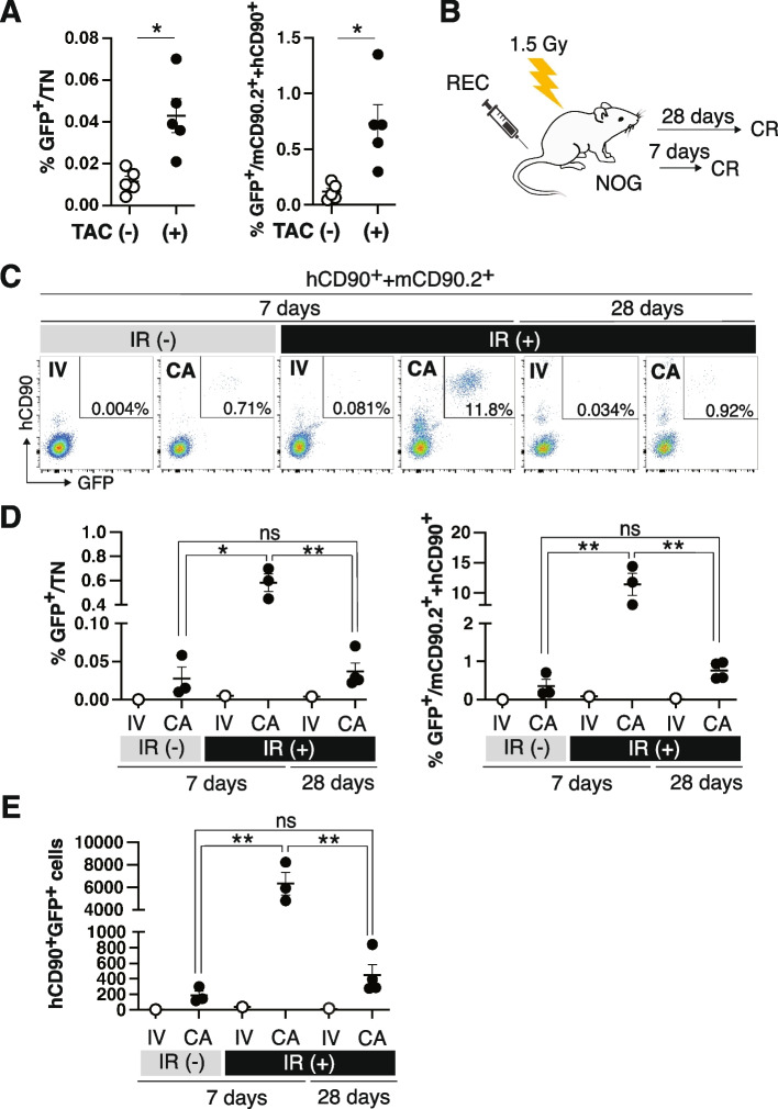
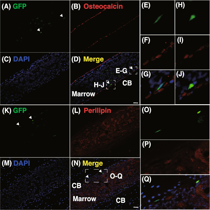
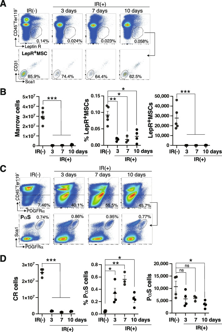
The transplantation of human mesenchymal stromal/stem cells (hMSCs) has potential as a curative and permanent therapy for congenital skeletal diseases. However, the self-renewal and differentiation capacities of hMSCs markedly vary. Therefore, cell proliferation and trilineage differentiation capacities were tested in vitro to characterize hMSCs before their clinical use. However, it remains unclear whether the ability of hMSCs in vitro accurately predicts that in living animals. The xenograft model is an alternative method for validating clinical MSCs. Nevertheless, the protocol still needs refinement, and it has yet to be established whether hMSCs, which are expanded in culture for clinical use, retain the ability to engraft and differentiate into adipogenic, osteogenic, and chondrogenic lineage cells in transplantation settings. In the present study, to establish a robust xenograft model of MSCs, we examined the delivery routes of hMSCs and the immunological state of recipients. The intra-arterial injection of hMSCs into X-ray-irradiated (IR) NOG, a severely immunodeficient mouse, achieved the highest engraftment but failed to sustain long-term engraftment. We demonstrated that graft cells localized to a collagenase-released fraction (CR), in which endogenous colony-forming cells reside. We also showed that Pdgfrα+Sca1+ MSCs (PαS), which reside in the CR fraction, resisted IR. These results show that our protocol enables hMSCs to fulfill a high level of engraftment in mouse bone marrow in the short term. In contrast, long-term reconstitution was restricted, at least partially, because of IR-resistant endogenous MSCs.
Keywords: Mesenchymal stem cells; Mesenchymal stromal cells; NOG; Regenerative medicine; Transplantation; Xenograft model.
© 2024. The Author(s).
Conflict of interest statement
The authors declare that they have no financial or non-financial competing interests.
Figures

Similar articles
-
Adipose tissue-derived human mesenchymal stromal cells can better suppress complement lysis, engraft and inhibit acute graft-versus-host disease in mice.Stem Cell Res Ther. 2023 Jun 25;14(1):167. doi: 10.1186/s13287-023-03380-x. Stem Cell Res Ther. 2023. PMID: 37357314 Free PMC article.
-
Isolation, characterization and the multi-lineage differentiation potential of rabbit bone marrow-derived mesenchymal stem cells.J Anat. 2013 Apr;222(4):437-50. doi: 10.1111/joa.12032. J Anat. 2013. PMID: 23510053 Free PMC article.
-
Large-Scale Automated Hollow-Fiber Bioreactor Expansion of Umbilical Cord-Derived Human Mesenchymal Stromal Cells for Neurological Disorders.Neurochem Res. 2020 Jan;45(1):204-214. doi: 10.1007/s11064-019-02925-y. Epub 2019 Dec 11. Neurochem Res. 2020. PMID: 31828497
-
Potential role of herbal remedies in stem cell therapy: proliferation and differentiation of human mesenchymal stromal cells.Stem Cell Res Ther. 2016 Aug 11;7(1):110. doi: 10.1186/s13287-016-0366-4. Stem Cell Res Ther. 2016. PMID: 27515026 Free PMC article. Review.
-
Senescence in Human Mesenchymal Stem Cells: Functional Changes and Implications in Stem Cell-Based Therapy.Int J Mol Sci. 2016 Jul 19;17(7):1164. doi: 10.3390/ijms17071164. Int J Mol Sci. 2016. PMID: 27447618 Free PMC article. Review.
Cited by
-
Perspectives and Limitations of Mesenchymal Stem Cell-Based Therapy for Corneal Injuries and Retinal Diseases.Cell Transplant. 2025 Jan-Dec;34:9636897241312798. doi: 10.1177/09636897241312798. Cell Transplant. 2025. PMID: 39856809 Free PMC article. Review.
References
-
- Dominici M, Le Blanc K, Mueller I, Slaper-Cortenbach I, Marini F, Krause D, Deans R, Keating A, Prockop DJ, Horwitz E. Minimal criteria for defining multipotent mesenchymal stromal cells. The International Society for Cellular Therapy position statement. Cytotherapy. 2006;8(4):315–7. 10.1080/14653240600855905. - PubMed
-
- Horwitz EM, Le Blanc K, Dominici M, Mueller I, Slaper-Cortenbach I, Marini FC, Deans RJ, Krause DS, Keating A. International society for cellular therapy. Clarification of the nomenclature for MSC: the international society for cellular therapy position statement. Cytotherapy. 2005;7(5):393–5. 10.1080/14653240500319234. - PubMed
-
- Taketani T, Oyama C, Mihara A, Tanabe Y, Abe M, Hirade T, Yamamoto S, Bo R, Kanai R, Tadenuma T, Michibata Y, Yamamoto S, Hattori M, Katsube Y, Ohnishi H, Sasao M, Oda Y, Hattori K, Yuba S, Ohgushi H, Yamaguchi S. Ex vivo expanded allogeneic mesenchymal stem cells with bone marrow transplantation improved osteogenesis in infants with severe hypophosphatasia. Cell Transplant. 2015;24(10):1931–43. 10.3727/096368914X68541. - PubMed
-
- Pittenger MF, Mackay AM, Beck SC, Jaiswal RK, Douglas R, Mosca JD, Moorman MA, Simonetti DW, Craig S, Marshak DR. Multilineage potential of adult human mesenchymal stem cells. Science. 1999;284(5411):143–7. 10.1126/science.284.5411.143. - PubMed
LinkOut - more resources
Full Text Sources
