

Diagnostic value of transvaginal contrast-enhanced ultrasound in identifying benign and malignant endometrial lesions and assessing myometrial invasion
- PMID: 39327718
- PMCID: PMC11532519
- DOI: 10.14366/usg.24097
Diagnostic value of transvaginal contrast-enhanced ultrasound in identifying benign and malignant endometrial lesions and assessing myometrial invasion
Abstract
Purpose: The objective of this study was to evaluate the diagnostic value of transvaginal contrastenhanced ultrasound (CEUS) in differentiating benign from malignant endometrial lesions and assessing the extent of myometrial invasion.
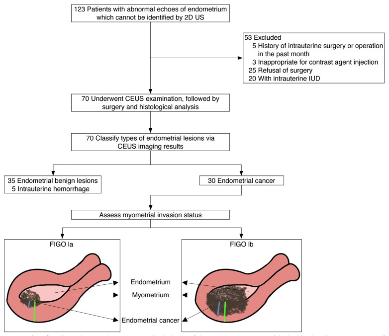
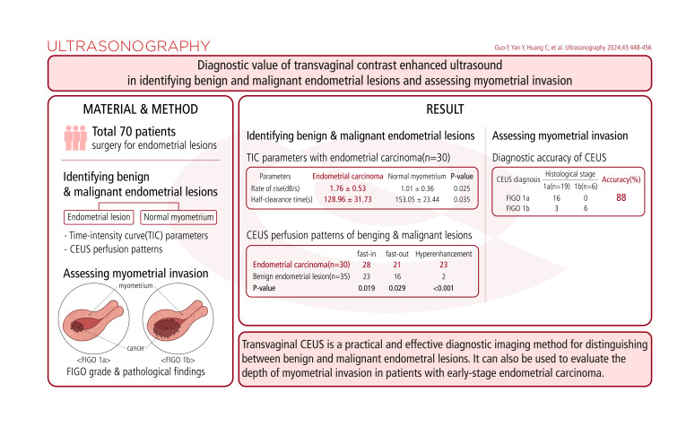
Methods: A total of 70 patients who underwent surgery for endometrial lesions at the authors' hospital were selected. Transvaginal ultrasound examination and CEUS were performed for quantitative and qualitative analysis. Based on the CEUS results, an International Federation of Gynecology and Obstetrics (FIGO) disease grade was assigned and compared with pathological findings.
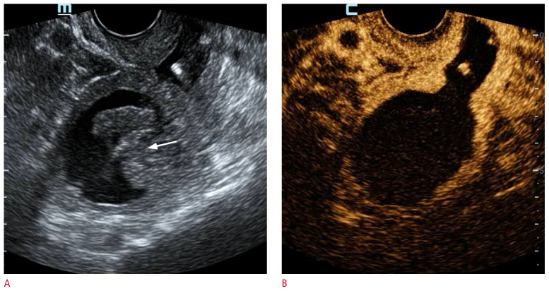
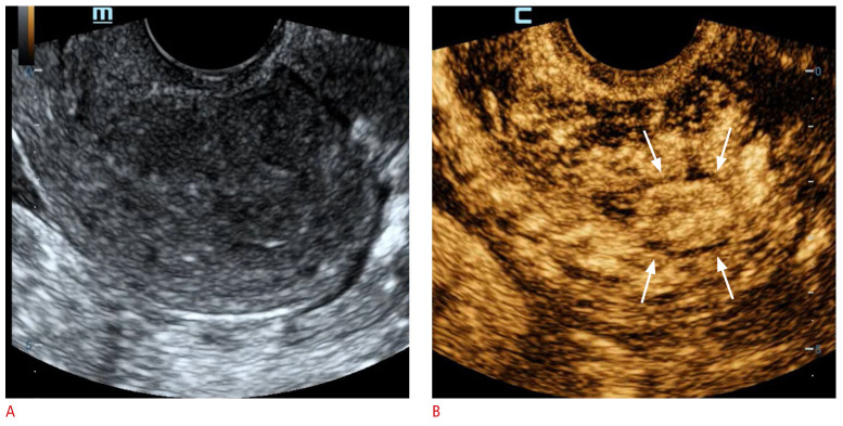
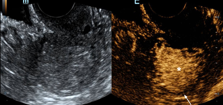
Results: Postmenopausal vaginal bleeding is a key clinical manifestation of endometrial carcinoma. Among the patients with endometrial carcinoma, compared with normal myometrium, the lesion areas exhibited a greater rate of rise (defined as enhanced intensity divided by enhancement time) and a shorter half-clearance time (P<0.05). These findings suggest that in endometrial carcinoma, the contrast agent displays a "fast-in/fast-out/hyperenhancement" perfusion pattern. In contrast, the characteristic perfusion pattern for benign endometrial lesions is low enhancement (P<0.05). The diagnostic accuracy of CEUS in detecting myometrial invasion was 88% (22 of 25 cases).
Conclusion: Transvaginal CEUS is a practical and effective diagnostic imaging method for distinguishing between benign and malignant endometrial lesions. It can also be used to evaluate the depth of myometrial invasion in patients with early-stage endometrial carcinoma.
Keywords: Contrast-enhanced ultrasound; Endometrial benign lesions; Endometrial neoplasms; Myometrial invasion.
Conflict of interest statement
No potential conflict of interest was reported by the authors.
Figures
Similar articles
-
Role of transvaginal contrast-enhanced ultrasound in the early diagnosis of endometrial carcinoma.Chin Med J (Engl). 2012 Feb;125(3):416-21. Chin Med J (Engl). 2012. PMID: 22490395
-
Application of Combined Two-Dimensional and Three-Dimensional Transvaginal Contrast Enhanced Ultrasound in the Diagnosis of Endometrial Carcinoma.Biomed Res Int. 2015;2015:292743. doi: 10.1155/2015/292743. Epub 2015 May 18. Biomed Res Int. 2015. PMID: 26090396 Free PMC article.
-
Estimating the depth of myometrial involvement by endometrial carcinoma: efficacy of transvaginal sonography vs MR imaging.AJR Am J Roentgenol. 1993 Mar;160(3):533-8. doi: 10.2214/ajr.160.3.8430547. AJR Am J Roentgenol. 1993. PMID: 8430547
-
[Assessment of myometrial invasion in endometrial cancer by transvaginal ultrasonography].Orv Hetil. 1997 May 25;138(21):1323-7. Orv Hetil. 1997. PMID: 9254351 Review. Hungarian.
-
[Diagnostic and preoperative staging of endometrial carcinoma with transvaginal sonography--a review].Zentralbl Gynakol. 2006 Oct;128(5):246-54. doi: 10.1055/s-2006-921420. Zentralbl Gynakol. 2006. PMID: 17001559 Review. German.
Cited by
-
Diagnostic value of contrast-enhanced ultrasound in endometrial lesions.BMC Womens Health. 2025 Jul 9;25(1):338. doi: 10.1186/s12905-025-03860-7. BMC Womens Health. 2025. PMID: 40634988 Free PMC article.
-
Diagnostic value of contrast-enhanced ultrasound for the depth of myometrial infiltration in early endometrial cancer: a meta-analysis.Front Oncol. 2025 Mar 7;15:1493246. doi: 10.3389/fonc.2025.1493246. eCollection 2025. Front Oncol. 2025. PMID: 40110205 Free PMC article.
References
-
- Crosbie EJ, Kitson SJ, McAlpine JN, Mukhopadhyay A, Powell ME, Singh N. Endometrial cancer. Lancet. 2022;399:1412–1428. - PubMed
-
- Mirza MR, Chase DM, Slomovitz BM, dePont Christensen R, Novak Z, Black D, et al. Dostarlimab for primary advanced or recurrent endometrial cancer. N Engl J Med. 2023;388:2145–2158. - PubMed
-
- Badea R, Ciobanu L. Contrast enhanced and Doppler ultrasonography in the characterization of the microcirculation: expectancies and performances. Med Ultrason. 2012;14:307–317. - PubMed
Grants and funding
LinkOut - more resources
Full Text Sources
