

Influencing factors of glymphatic system during perioperative period
- PMID: 39328423
- PMCID: PMC11424614
- DOI: 10.3389/fnins.2024.1428085
Influencing factors of glymphatic system during perioperative period
Abstract
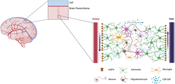
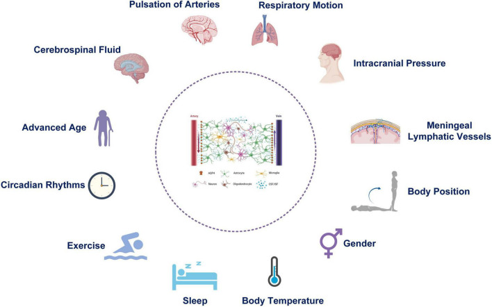
The glymphatic system is a functional cerebrospinal fluid circulatory system that uses peri-arterial space for inflow of cerebrospinal fluid and peri-venous space for efflux of cerebrospinal fluid from brain parenchyma. This brain-wide fluid transport pathway facilitates the exchange between cerebrospinal fluid and interstitial fluid and clears metabolic waste from the metabolically active brain. Multiple lines of work show that the glymphatic system is crucial to normal brain functions, and the dysfunction of the glymphatic system is closely associated with various neurological disorders, including aging, neurodegeneration, and acute brain injury. Currently, it is common to explore the functional and molecular mechanisms of the glymphatic system based on animal models. The function of glymphatic system during perioperative period is affected by many factors such as physiological, pathological, anesthetic and operative methods. To provide a reference for the interpretation of the results of glymphatic system studies during perioperative period, this article comprehensively reviews the physiological and pathological factors that interfere with the function of the glymphatic system during perioperative period, investigates the effects of anesthetic drugs on glymphatic system function and the potential underlying mechanisms, describes operative methods that interfere with the function of the glymphatic system, and potential intervention strategies based on the glymphatic system. Future, these variables should be taken into account as critical covariates in the design of functional studies on the glymphatic system.
Keywords: AQP4; anesthetic drugs; cerebrospinal fluid; glymphatic system; pathology; perioperative; physiology.
Copyright © 2024 Dong, Liu, Han, Wang, Jiang, Wang and Gu.
Conflict of interest statement
The authors declare that the research was conducted in the absence of any commercial or financial relationships that could be construed as a potential conflict of interest.
Figures
Similar articles
-
The Glymphatic System (En)during Inflammation.Int J Mol Sci. 2021 Jul 13;22(14):7491. doi: 10.3390/ijms22147491. Int J Mol Sci. 2021. PMID: 34299111 Free PMC article. Review.
-
Glymphatic system: an emerging therapeutic approach for neurological disorders.Front Mol Neurosci. 2023 Jul 6;16:1138769. doi: 10.3389/fnmol.2023.1138769. eCollection 2023. Front Mol Neurosci. 2023. PMID: 37485040 Free PMC article. Review.
-
Loss of aquaporin-4 results in glymphatic system dysfunction via brain-wide interstitial fluid stagnation.Elife. 2023 Feb 9;12:e82232. doi: 10.7554/eLife.82232. Elife. 2023. PMID: 36757363 Free PMC article.
-
Glymphatic System in the Central Nervous System, a Novel Therapeutic Direction Against Brain Edema After Stroke.Front Aging Neurosci. 2021 Aug 4;13:698036. doi: 10.3389/fnagi.2021.698036. eCollection 2021. Front Aging Neurosci. 2021. PMID: 34421575 Free PMC article.
-
Gadolinium-based Contrast Media, Cerebrospinal Fluid and the Glymphatic System: Possible Mechanisms for the Deposition of Gadolinium in the Brain.Magn Reson Med Sci. 2018 Apr 10;17(2):111-119. doi: 10.2463/mrms.rev.2017-0116. Epub 2018 Jan 25. Magn Reson Med Sci. 2018. PMID: 29367513 Free PMC article.
Cited by
-
Alzheimer's disease and insomnia: a bibliometric study and visualization analysis.Front Aging Neurosci. 2025 Apr 8;17:1542607. doi: 10.3389/fnagi.2025.1542607. eCollection 2025. Front Aging Neurosci. 2025. PMID: 40264463 Free PMC article. Review.
References
-
- Abdul Y., Li W., Ward R., Abdelsaid M., Hafez S., Dong G., et al. (2021). Deferoxamine treatment prevents post-stroke vasoregression and neurovascular unit remodeling leading to improved functional outcomes in type 2 male diabetic rats: Role of endothelial ferroptosis. Transl. Stroke Res. 12 615–630. 10.1007/s12975-020-00844-7 - DOI - PMC - PubMed
Publication types
LinkOut - more resources
Full Text Sources