

Jejunal amyloidosis: a rare case presented with sudden weight loss and altered bowel habits
- PMID: 39328977
- PMCID: PMC11422502
- DOI: 10.1093/bjrcr/uaae025
Jejunal amyloidosis: a rare case presented with sudden weight loss and altered bowel habits
Abstract
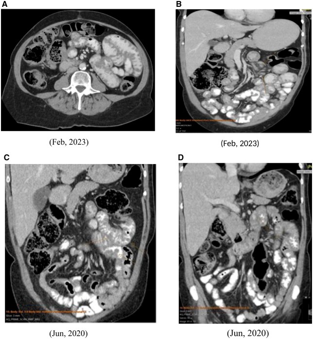
Amyloidosis is a multisystem disease characterized by the extracellular deposition of abnormal protein fibrils in various tissues and organs. It is a systemic disease mainly affecting the kidneys, liver, and spleen. In the GI tract, the duodenum is most commonly involved, followed by the stomach. We came across a relatively unusual case of jejunal amyloidosis, where the presentation was with weight loss, abdominal pain, and changes in bowel habits. Radiological findings were non-specific, such as thickening of small bowel loops and small nodular bowel wall lesions, etc. Endoscopic features were also not characteristic of any particular disease entity. Therefore, it was hard to narrow down the differential diagnosis based on endoscopy and clinic-radiological grounds. The diagnosis remained a mystery until the outcome of the final histopathology report. This case study will facilitate readers in considering this entity in the differential list if they encounter mimicking clinical and/or radiological features.
© Crown copyright 2024.
Conflict of interest statement
None declared.
Figures


Similar articles
-
Vesicoureteral Reflux.2024 Apr 30. In: StatPearls [Internet]. Treasure Island (FL): StatPearls Publishing; 2025 Jan–. 2024 Apr 30. In: StatPearls [Internet]. Treasure Island (FL): StatPearls Publishing; 2025 Jan–. PMID: 33085409 Free Books & Documents.
-
Primary gastrointestinal amyloidosis with gastrointestinal hemorrhage and intestinal pseudo-obstruction: a report of a rare case.Clin J Gastroenterol. 2019 Jun;12(3):258-262. doi: 10.1007/s12328-018-00929-9. Epub 2018 Dec 20. Clin J Gastroenterol. 2019. PMID: 30574660 Free PMC article.
-
[Gastrointestinal hemorrhage as an uncommon form of presentation of primary intestinal amyloidosis: case report].Rev Gastroenterol Peru. 2023 Oct-Dec;43(4):358-363. Rev Gastroenterol Peru. 2023. PMID: 38228302 Spanish.
-
Intestinal amyloidosis: Clinical manifestations and diagnostic challenge.Adv Clin Exp Med. 2021 May;30(5):563-570. doi: 10.17219/acem/133521. Adv Clin Exp Med. 2021. PMID: 33974753 Review.
-
Amyloid and the GI tract.Expert Rev Gastroenterol Hepatol. 2009 Dec;3(6):615-30. doi: 10.1586/egh.09.59. Expert Rev Gastroenterol Hepatol. 2009. PMID: 19929583 Review.
References
-
- Bustamante JG, Zaidi SRH.. Amyloidosis. In: Bustamante JG, Zaidi SRH, eds. StatPearls. StatPearls Publishing; 2023.
-
- Kaiserling E, Kröber S.. Massive intestinal hemorrhage associated with intestinal amyloidosis. An investigation of underlying pathologic processes. Gen Diagn Pathol. 1995;141(2):147-154. - PubMed
Publication types
LinkOut - more resources
Full Text Sources
