. 2024 Dec;30(6):218.
doi: 10.3892/mmr.2024.13342. Epub 2024 Sep 27.

Therapeutic role of histone deacetylase inhibition in an in vitro model of Graves' orbitopathy

Affiliations

Therapeutic role of histone deacetylase inhibition in an in vitro model of Graves' orbitopathy

Hyeong Ju Byeon et al. Mol Med Rep. 2024 Dec.

Abstract

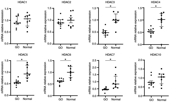
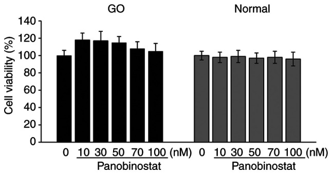
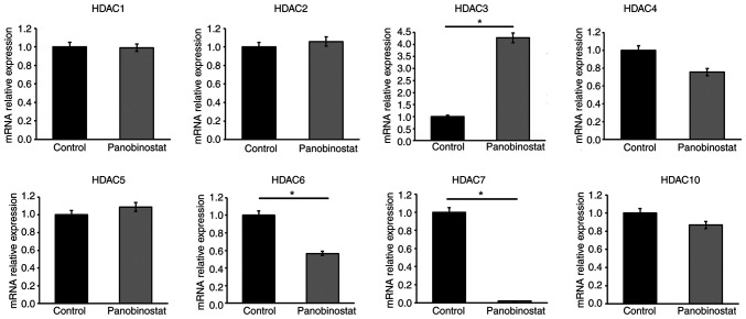
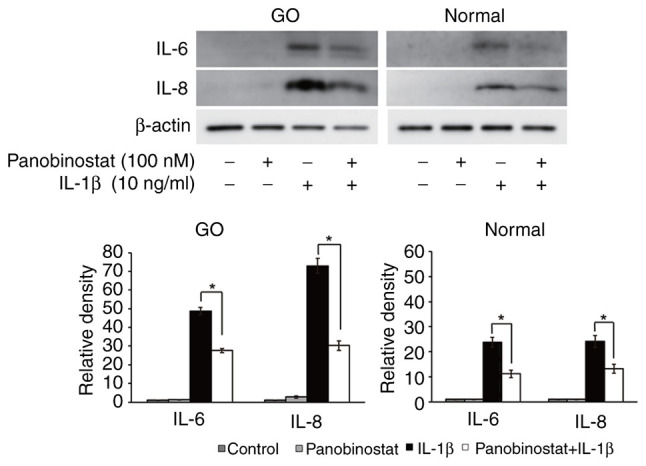
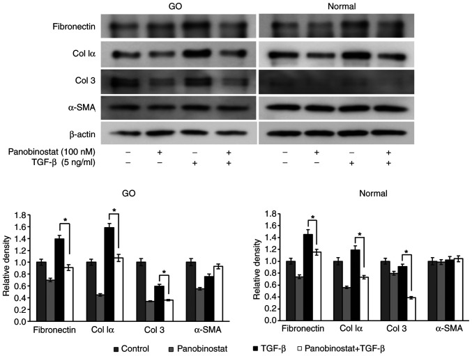
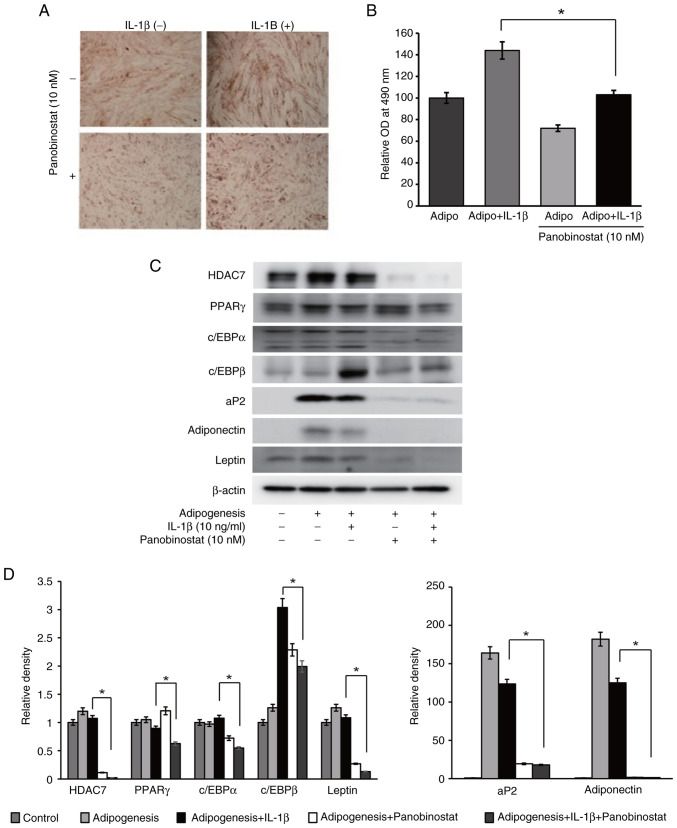
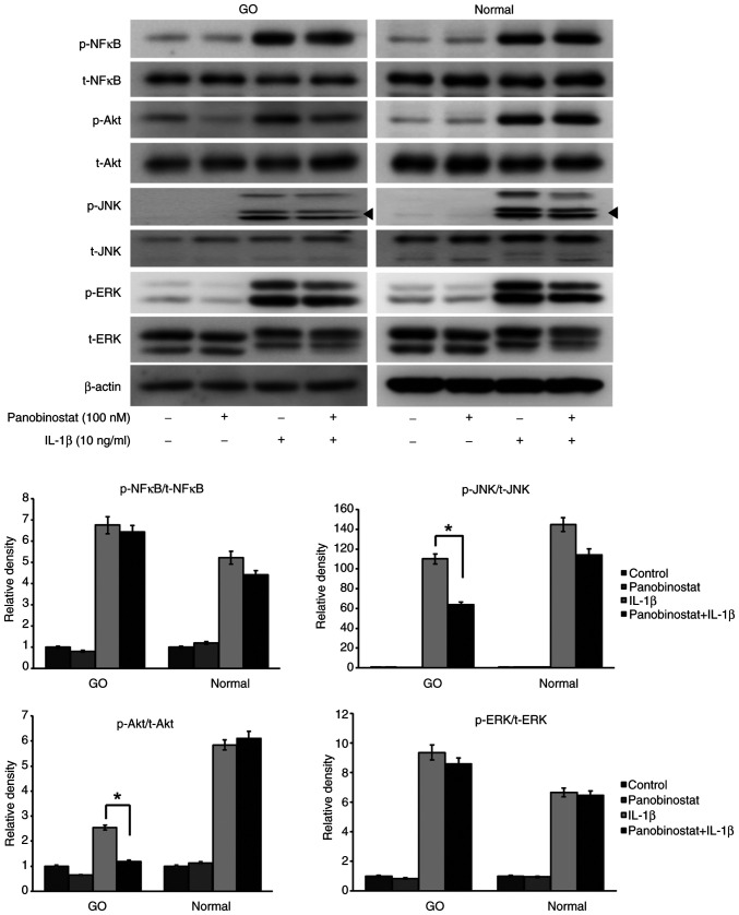
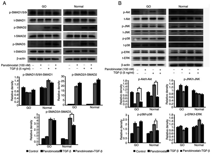
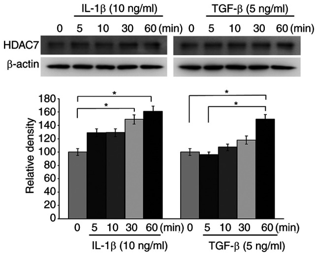
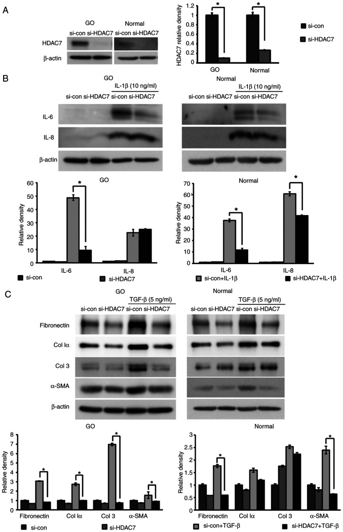
Graves' orbitopathy (GO), a manifestation of Graves' disease, is characterized by orbital fibroblast‑induced inflammation, leading to fibrosis or adipogenesis. Histone deacetylase (HDAC) serves a central role in autoimmune diseases and fibrosis. The present study investigated HDAC inhibition in orbital fibroblasts from patients with GO to evaluate its potential as a therapeutic agent. Primary cultured orbital fibroblasts were treated with an HDAC inhibitor, panobinostat, under the stimulation of IL‑1β, TGF‑β or adipogenic medium. Inflammatory cytokines, and fibrosis‑ and adipogenesis‑related proteins were analyzed using western blotting. The effects of panobinostat on HDAC mRNA expression were measured in GO orbital fibroblasts, and specific HDACs were inhibited using small interfering RNA transfection. Panobinostat significantly reduced the IL‑1β‑induced production of inflammatory cytokines and TGF‑β‑induced production of fibrosis‑related proteins. It also suppressed adipocyte differentiation and adipogenic transcription factor production. Furthermore, it significantly attenuated HDAC7 mRNA expression in GO orbital fibroblasts. In addition, the silencing of HDAC7 led to anti‑inflammatory and anti‑fibrotic effects. In conclusion, by inhibiting HDAC7 gene expression, panobinostat may suppress the production of inflammatory cytokines, profibrotic proteins and adipogenesis in GO orbital fibroblasts. The present in vitro study suggested that HDAC7 could be a potential therapeutic target for inhibiting the inflammatory, adipogenic and fibrotic mechanisms of GO.

Keywords: Graves' orbitopathy; histone deacetylase 7; histone deacetylase inhibitor; orbital fibroblast; panobinostat.


Conflict of interest statement

The authors declare that they have no competing interests.

Figures

Figure 1.
Figure 1.
HDAC mRNA expression in GO and control orbital tissues. Orbital tissues from patients with GO (n=10) and controls (n=8) were analyzed to evaluate the mRNA expression levels of HDACs (class I: HDAC1, 2 and 3; class IIa: HDAC4, 5 and 7; class IIb: HDAC6 and 10). Reverse transcription-quantitative PCR was performed, and the results are presented as the median and interquartile ranges compared with normal controls. Statistical significance was determined using Mann-Whitney U-test. *P<0.05 vs. normal control. GO, Graves' orbitopathy; HDAC, histone deacetylase.
Figure 2.
Figure 2.
Cell viability after treatment with panobinostat. Orbital fibroblasts from patients with GO (n=3) and controls (n=3) were seeded in 24-well culture plates and treated with various concentrations of panobinostat (control, 10, 30, 50, 70 and 100 nM) for 24 h. The MTT assay was repeated twice for cells from 3 different individuals. Results are presented as the mean ± SD relative to the untreated control. GO, Graves' orbitopathy.
Figure 3.
Figure 3.
Effect of panobinostat on the mRNA expression levels of HDACs. Orbital fibroblasts from patients with GO (n=3) were cultured with panobinostat (100 nM) for 24 h. GO orbital fibroblasts cultured without panobinostat for 24 h were used as controls. mRNA expression levels of HDACs (class I: HDAC1, 2 and 3; class IIa: HDAC4, 5 and 7; class IIb: HDAC6 and 10) were measured by reverse transcription-quantitative PCR. All experiments were conducted twice on samples from 3 individuals. Results are presented as the mean ± SD relative to the control. Statistical significance was determined using Mann-Whitney U-test. *P<0.05 vs. untreated control. GO, Graves' orbitopathy; HDAC, histone deacetylase.
Figure 4.
Figure 4.
Suppressive effect of panobinostat on proinflammatory cytokine expression. Orbital fibroblasts from patients with GO (n=3) and controls (n=3) were pretreated with panobinostat (100 nM) for 30 min and then stimulated with IL-1β (10 ng/ml) for 24 h. Western blotting was performed to examine the protein expression levels of the proinflammatory cytokines IL-6 and IL-8. Representative gel images are shown. Densitometry was performed and levels were normalized to β-actin in the same sample. All experiments were conducted twice on samples from three individuals. Data are presented as the mean ± SD. Statistical significance was determined using Mann-Whitney U-test to compare the effect of panobinostat under IL-1β stimulation. *P<0.05. GO, Graves' orbitopathy.
Figure 5.
Figure 5.
Suppressive effect of panobinostat on the expression of TGF-β-induced profibrotic proteins. Orbital fibroblasts from patients with GO (n=3) and controls (n=3) were pretreated with panobinostat (100 nM) for 30 min and then stimulated with TGF-β (5 ng/ml) for 24 h. The expression levels of the profibrotic proteins fibronectin, Col Iα, Col 3 and α-SMA were determined by western blot analysis. Representative gel images are shown. β-actin was used for normalization. All experiments were conducted twice on samples from three individuals. Data are presented as the mean ± SD. Statistical significance was determined using Mann-Whitney U-test to compare the effect of panobinostat under TGF-β stimulation. *P<0.05. α-SMA, α-smooth muscle actin; Col, collagen; GO, Graves' orbitopathy.
Figure 6.
Figure 6.
Suppressive effect of panobinostat on adipogenesis. Graves' orbitopathy orbital fibroblasts (n=3) were differentiated into adipocytes after 14 days of incubation in adipogenic medium. Panobinostat (10 nM) and/or IL-1β (10 ng/ml) was added during the adipogenesis. All experiments were performed twice. (A) To evaluate adipocyte differentiation, cells were stained with Oil Red O, and cytoplasmic lipid droplets were examined under a microscope (magnification, ×40). (B) Stained cell lysates were solubilized, and absorbance was measured using a spectrophotometer at 490 nm. Data are presented as the mean ± SD. Statistical significance was determined using Mann-Whitney U-test to compare the effect of panobinostat under adipogenic stimulation with IL-1β. *P<0.05. (C) Adipogenic transcription factors, including PPARγ, c/EBPα, c/EBPβ, aP2, adiponectin and leptin, and HDAC7 were analyzed by western blotting after 14 days of adipogenic differentiation of orbital fibroblasts. Representative gel images are shown. (D) Densitometric analysis results of western blotting. Data are presented as the mean ± SD normalized to β-actin. Statistical significance was determined using Mann-Whitney U-test to compare the effect of panobinostat under adipogenic stimulation with IL-1β. *P<0.05. aP2, adipocyte protein 2; c/EBP, CCAAT-enhancer-binding protein; HDAC7, histone deacetylase 7; OD, optical density; PPARγ, peroxisome proliferator-activated receptor γ.
Figure 7.
Figure 7.
Effect of panobinostat on signal proteins under IL-1β stimulation. Orbital fibroblasts from patients with GO (n=3) and controls (n=3) were pretreated with panobinostat (100 nM) for 24 h, followed by IL-1β (10 ng/ml) treatment for 15 min. The t- and p-NF-κB, Akt, JNK and ERK proteins were analyzed through western blotting. Representative gel images are shown. Arrow indicates the p-JNK bands. β-actin was used as a loading control. After semi-quantification using densitometry, the ratio of p-/t-proteins was measured and compared with that of the control. Data are presented as the mean ± SD. Statistical significance was determined using Mann-Whitney U-test to compare the effect of panobinostat under IL-1β stimulation. *P<0.05. GO, Graves' orbitopathy; p-, phosphorylated; t-, total.
Figure 8.
Figure 8.
Effect of panobinostat on signal proteins under TGF-β stimulation. (A) Orbital fibroblasts were exposed to 100 nM panobinostat for 3 h, and then stimulated with TGF-β (5 ng/ml) for 1 h. The p-form of the SMAD signaling transducers, SMAD1/5/9, SMAD2 and SMAD3, were analyzed by western blotting, as were the total forms of SMAD1, SMAD2 and SMAD3. (B) Non-SMAD pathways under TGF-β (5 ng/ml) stimulation for 1 h were examined in orbital fibroblasts. Orbital fibroblasts were pretreated with 100 nM panobinostat for 3 h before TGF-β treatment. Western blot analysis of p- and t-Akt, JNK, p38 and ERK non-SMAD pathway molecules was performed. Orbital fibroblasts from three different GO samples and healthy subjects were used in the present study. β-actin was used as a loading control. The ratio of p-/t-proteins was measured and compared with that of the control, and data are presented as the mean ± SD. Statistical significance was determined using Mann-Whitney U-test to compare the effect of panobinostat under TGF-β stimulation. *P<0.05. GO, Graves' orbitopathy; p-, phosphorylated; t-, total.
Figure 9.
Figure 9.
HDAC7 expression under stimulation with IL-1β and TGF-β in GO orbital fibroblasts. Orbital fibroblasts from patients with GO (n=3) were stimulated with IL-1β (10 ng/ml) and TGF-β (5 ng/ml) for increasing time intervals (0–60 min). HDAC7 release was measured at the indicated times (0, 5, 10, 30 and 60 min) through western blot analysis. Representative gel images are shown. β-actin was used as a loading control. All experiments were conducted twice on samples from 3 individuals. Data measured by densitometry are presented as the mean ± SD. Relative density was determined with respect to the 0-min time point. Each group was compared using the Kruskal-Wallis test followed by Dunn's post hoc test. *P<0.05. GO, Graves' orbitopathy; HDAC7, histone deacetylase 7.
Figure 10.
Figure 10.
Anti-inflammatory and anti-fibrotic effects of silencing HDAC7 in orbital fibroblasts. HDAC7 was knocked down via transfection with siRNA (10 nM) for 24 h and cells were maintained for 48 h. (A) Silencing efficiency of si-HDAC7 was demonstrated in both GO and normal orbital fibroblasts (n=3) compared with si-con. (B) Cells were stimulated with IL-1β (10 ng/ml) for 24 h. Western blot analysis was performed to investigate the expression levels of the proinflammatory cytokines IL-6 and IL-8. (C) Orbital fibroblasts were treated with TGF-β (5 ng/ml) for 24 h, and profibrotic proteins, including fibronectin, Col Iα, Col 3 and α-SMA, were examined using western blot analysis. β-actin was used as a loading control for normalization. Each experiment was performed in duplicate. Data are presented as the mean ± SD. Statistical significance was determined using Mann-Whitney U-test to compare the effect of silencing HDAC7 under IL-1β or TGF-β stimulation. *P<0.05. α-SMA, α-smooth muscle actin; Col, collagen; con, control; GO, Graves' orbitopathy; HDAC7, histone deacetylase 7; si/siRNA, small interfering RNA.

Similar articles

References

    1. Lazarus JH. Epidemiology of Graves' orbitopathy (GO) and relationship with thyroid disease. Best Pract Res Clin Endocrinol Metab. 2012;26:273–279. doi: 10.1016/j.beem.2011.10.005. - DOI - PubMed
    1. Bahn RS. Graves' ophthalmopathy. N Engl J Med. 2010;362:726–738. doi: 10.1056/NEJMra0905750. - DOI - PMC - PubMed
    1. Bartley GB, Fatourechi V, Kadrmas EF, Jacobsen SJ, Ilstrup DM, Garrity JA, Gorman CA. Clinical features of Graves' ophthalmopathy in an incidence cohort. Am J Ophthalmol. 1996;121:284–290. doi: 10.1016/S0002-9394(14)70276-4. - DOI - PubMed
    1. Rotondo Dottore G, Torregrossa L, Lanzolla G, Mariotti S, Menconi F, Piaggi P, Cristofani Mencacci L, Posarelli C, Maglionico MN, Dallan I, et al. Role of the mononuclear cell infiltrate in Graves' orbitopathy (GO): Results of a large cohort study. J Endocrinol Invest. 2022;45:563–572. doi: 10.1007/s40618-021-01692-4. - DOI - PubMed
    1. Huang Y, Fang S, Li D, Zhou H, Li B, Fan X. The involvement of T cell pathogenesis in thyroid-associated ophthalmopathy. Eye (Lond) 2019;33:176–182. doi: 10.1038/s41433-018-0279-9. - DOI - PMC - PubMed

MeSH terms