

Oxygen matters: Unraveling the role of oxygen in the neuronal response to cisplatin
- PMID: 39329299
- PMCID: PMC11625991
- DOI: 10.1111/jns.12659
Oxygen matters: Unraveling the role of oxygen in the neuronal response to cisplatin
Abstract
Background and aims: Cell culture is a fundamental experimental tool for understanding cell physiology. However, translating these findings to in vivo settings has proven challenging. Replicating donor tissue conditions, including oxygen levels, is crucial for achieving meaningful results. Nevertheless, oxygen culture conditions are often overlooked, particularly in the context of chemotherapy-induced neurotoxicity.
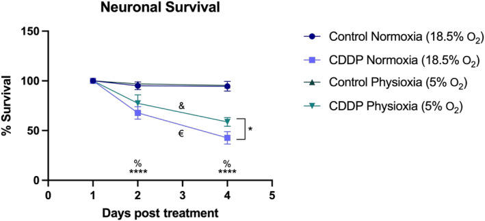
Methods: In this study, we investigated the role of oxygen levels in primary neuronal cultures by comparing neuronal performance under cisplatin exposure (1 μg/mL) in supraphysiological normoxia (representing atmospheric conditions in a standard incubator; 18.5% O2) and physioxia (representing physiologic oxygen conditions in nervous tissue; 5% O2). Experiments were also conducted to assess survival, neurite development, senescence marker expression, and proinflammatory cytokine secretion.
Results: Under control conditions, both oxygen concentration conditions exhibited similar behaviors. However, after cisplatin administration, sensory neurons cultured under supraphysiological normoxic conditions show higher mortality, exhibit an evolutionarily proinflammatory cytokine profile over time, and activate apoptotic-regulated neuron death markers. In contrast, under physiological conditions, neurons treated with cisplatin exhibited senescence marker expression and an attenuated inflammatory secretome.
Interpretation: These results underscore the critical role of oxygen in neuronal culture, particularly in studying compounds where neuronal damage is mechanistically linked to oxidative stress. Even at identical doses of evaluated neurotoxic drugs, distinct cellular phenotypic fates can emerge, impacting translatability to the in vivo setting.
Keywords: cell culture; chemotherapy‐induced neuropathy; cisplatin; neurotoxicity; oxygen.
© 2024 The Author(s). Journal of the Peripheral Nervous System published by Wiley Periodicals LLC on behalf of Peripheral Nerve Society.
Conflict of interest statement
The authors declare no conflicts of interest.
Figures


Similar articles
-
Thymoquinone prevents cisplatin neurotoxicity in primary DRG neurons.Neurotoxicology. 2018 Dec;69:68-76. doi: 10.1016/j.neuro.2018.09.001. Epub 2018 Sep 15. Neurotoxicology. 2018. PMID: 30227172
-
Is 8% O2 more normoxic than 21% O2 for long-term in vitro cultures of human primary term cytotrophoblasts?Mol Hum Reprod. 2018 Apr 1;24(4):211-220. doi: 10.1093/molehr/gax069. Mol Hum Reprod. 2018. PMID: 29534204
-
Oxidative DNA Damage and Cisplatin Neurotoxicity Is Exacerbated by Inhibition of OGG1 Glycosylase Activity and APE1 Endonuclease Activity in Sensory Neurons.Int J Mol Sci. 2022 Feb 8;23(3):1909. doi: 10.3390/ijms23031909. Int J Mol Sci. 2022. PMID: 35163831 Free PMC article.
-
[The Effects of Cysplatin on Human Adipose Tissue Derived Mesenchymal Stromal Cells Under Different Oxygen Levels].Vestn Ross Akad Med Nauk. 2016;(2):114-20. doi: 10.15690/vramn614. Vestn Ross Akad Med Nauk. 2016. PMID: 27522712 Russian.
-
Supraphysiological Oxygen Levels in Mammalian Cell Culture: Current State and Future Perspectives.Cells. 2022 Oct 4;11(19):3123. doi: 10.3390/cells11193123. Cells. 2022. PMID: 36231085 Free PMC article. Review.
References
-
- Seyhan AA. Lost in translation: the valley of death across preclinical and clinical divide—identification of problems and overcoming obstacles. Transl Med Commun. 2019;4(1):1‐19. doi:10.1186/S41231-019-0050-7 - DOI
-
- Staff NP, Fehrenbacher JC, Caillaud M, Damaj MI, Segal RA, Rieger S. Pathogenesis of paclitaxel‐induced peripheral neuropathy: a current review of in vitro and in vivo findings using rodent and human model systems. Exp Neurol. 2020;324:113121. doi:10.1016/J.EXPNEUROL.2019.113121 - DOI - PMC - PubMed
MeSH terms
Substances
Grants and funding
LinkOut - more resources
Full Text Sources
