Review
. 2024 Dec;13(1):2403380.
doi: 10.1080/21623945.2024.2403380. Epub 2024 Sep 27.

Involvement of a battery of investigated genes in lipid droplet pathophysiology and associated comorbidities

Affiliations
Review

Involvement of a battery of investigated genes in lipid droplet pathophysiology and associated comorbidities

Sami N Al Harake et al. Adipocyte. 2024 Dec.

Abstract

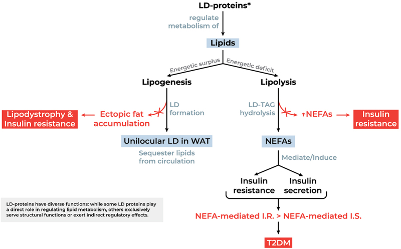
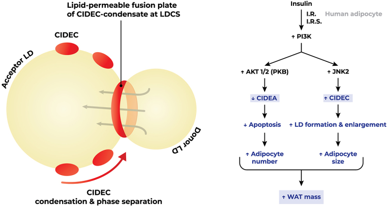
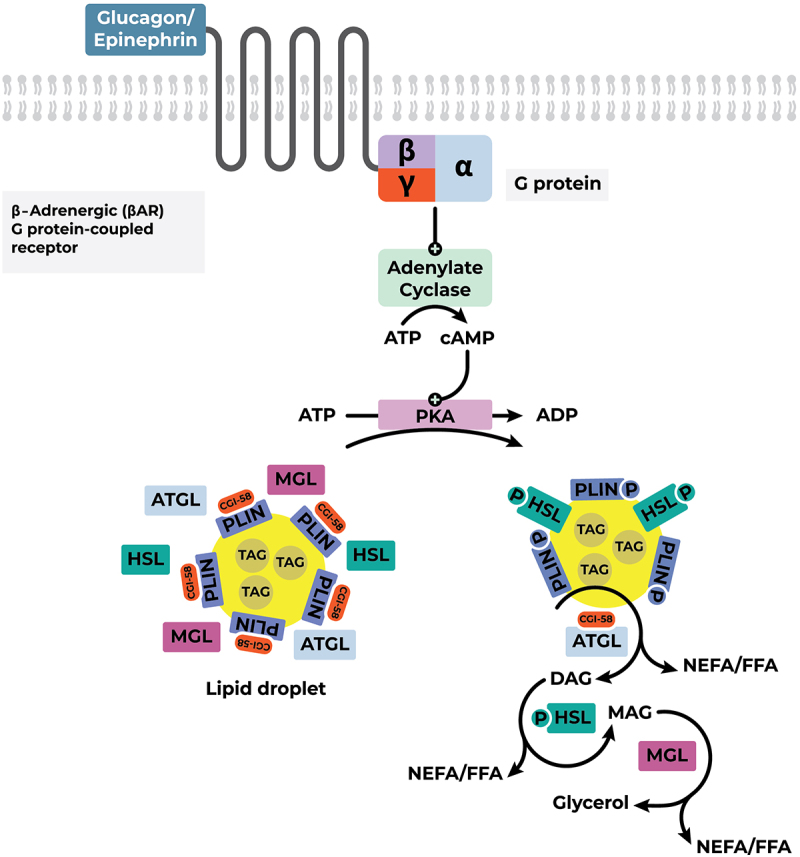
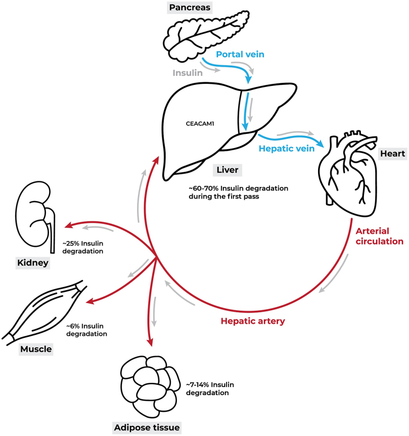
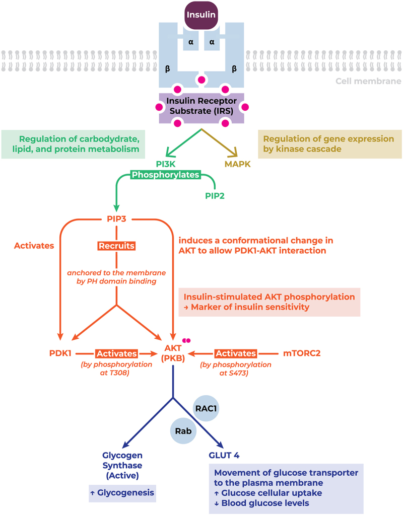
Lipid droplets (LDs) are highly specialized energy storage organelles involved in the maintenance of lipid homoeostasis by regulating lipid flux within white adipose tissue (WAT). The physiological function of adipocytes and LDs can be compromised by mutations in several genes, leading to NEFA-induced lipotoxicity, which ultimately manifests as metabolic complications, predominantly in the form of dyslipidemia, ectopic fat accumulation, and insulin resistance. In this review, we delineate the effects of mutations and deficiencies in genes - CIDEC, PPARG, BSCL2, AGPAT2, PLIN1, LIPE, LMNA, CAV1, CEACAM1, and INSR - involved in lipid droplet metabolism and their associated pathophysiological impairments, highlighting their roles in the development of lipodystrophies and metabolic dysfunction.

Keywords: Lipid droplet formation; adipogenesis; insulin resistance; lipid droplet hydrolysis; lipodystrophy.


Conflict of interest statement

No potential conflict of interest was reported by the author(s).

Figures

This schematic diagram describes the regulation of lipid metabolism by LD proteins. The diagram is divided into two main pathways: lipogenesis and lipolysis. Lipogenesis leads to unilocular lipid droplets in white adipose tissue, sequestering lipids from circulation. Lipolysis results in the release of non-esterified fatty acids (NEFAs). Impairments in these pathways mediate insulin resistance and contribute to the development of type 2 diabetes mellitus (T2DM), lipodystrophy and ectopic fat accumulation.
Figure 1.
Functional significance of LD-proteins in lipid metabolism and potential aberrations. NEFAs mediate insulin resistance by hindering insulin-induced glucose uptake and glycogen synthesis. Simultaneously, NEFAs drive glucose-stimulated insulin secretion. This nuance is crucial, as it implies that insulin resistance would not necessarily lead to diabetes as long as NEFA-mediated insulin secretion is able to compensate for NEFA-induced insulin resistance [5,6]. Abbreviations: I.R., insulin resistance; I.S., insulin secretion; LD, lipid droplet; LD-proteins, lipid droplet associated proteins; NEFA, non-esterified (free) fatty acids (plasma-circulating); T2DM, type 2 diabetes Mellitus; TAG, Triacylglycerol; WAT, white adipose tissue.
This figure illustrates the process of lipid droplet fusion mediated by CIDEC in adipocytes. On the left, CIDEC facilitates the fusion of a donor lipid droplet with an acceptor lipid droplet, involving condensation and phase separation at the lipid droplet contact site (LDCS), resulting in LD enlargement. On the right, insulin signalling activates PI3K, leading to two pathways: AKT activation increases CIDEA, reducing apoptosis and increasing adipocyte number, while JNK2 activation increases CIDEC, promoting LD formation and enlargement, leading to increased adipocyte size. Both pathways converge to an increase in white adipose tissue (WAT) mass.
Figure 2.
Insulin signalling triggers lipid droplet fusion. Insulin-induced anti-apoptosis and lipid droplet formation by differential regulation of CIDEA and CIDEC via Akt1/2 and JNK2-dependent pathways, respectively, in human adipocytes. CIDEC mediates lipid droplet fusion via gel-like condensation on lipid droplet cell surface. Figure readapted from [24] and [26]. Abbreviations: AKT/PKB, protein kinase B; I.R., insulin receptor; I.R.S., insulin receptor substrate; JNK2, c-Jun N-terminal kinase 2; LD, lipid droplet; LDCS, lipid droplet contact site; PI3K, phosphatidylinositol 3-kinase; WAT, white adipose tissue.
This figure depicts the signalling pathway conducing to the activation of lipolysis in lipid droplets. The diagram illustrates the activation of adenylate cyclase by a β-adrenergic G protein-coupled receptor, leading to the conversion of ATP to cAMP, which then activates protein kinase A (PKA). PKA phosphorylates Perilipin 1 (PLIN1) and hormone-sensitive lipase (HSL), facilitating the breakdown of triglycerides into glycerol and non-esterified fatty acids (NEFA/FFA).
Figure 3.
Mobilization of TAGs in lipid droplets of white adipose tissue. Abbreviations: ADP, Adenosine diphosphate; ATGL, adipose triglyceride lipase; ATP, Adenosine triphosphate; cAMP, cyclic Adenosine monophosphate; CGI-58, comparative gene identification-58 (co-activator of ATGL); DAG, Diacylglycerol; FFA, free fatty Acids; HSL, hormone sensitive lipase; MAG, Monoacylglycerol; MGL, monoglyceride lipase; NEFA, non-esterified fatty acids; PKA, protein kinase A; PLIN, perilipin-1; TAG, Triacylglycerol.
This diagram depicts the degradation of insulin in different tissues. Insulin is released by the pancreas and travels through the portal vein to reach the liver where it is primarily degraded. It then enters systemic circulation via the hepatic vein and is subsequently utilized by peripheral organs, such as the kidneys, muscles, and adipocytes.
Figure 4.
The journey of insulin. Insulin is discharged from the pancreas into the portal vein in a pulsatile manner, and it is primarily supplied to hepatocytes. The liver is the first organ to receive insulin, and it clears most of the insulin during the first passage, which accounts for around 60–70% of the insulin. The lingering insulin enters the systemic circulation, where peripheral tissues such as muscles, adipose tissue, and kidneys utilize it moderately, and then the liver degrades it again during the second passage through the hepatic artery. Modified from [149].
This diagram illustrates the insulin signalling pathway. Insulin binding to its receptor initiates the PI3K and MAPK signalling cascades. signalling downstream of the PI3K pathway mediates AKT phosphorylation which in turn facilitates the translocation of GLUT4 to the plasma membrane, increasing glucose uptake, and stimulates glycogen synthesis by activating glycogen synthase. This pathway regulates carbohydrate, lipid, and protein metabolism.
Figure 5.
INSR structure, downstream signalling pathway, and function. Upon Upon binding to its cognate receptor, insulin triggers the phosphorylation and recruitment of key signalling mediators conducive to their activation, thereby enhancing cellular glucose uptake, and promoting glycogenesis. Abbreviations: AKT/PKB, protein kinase B (serine/threonine-specific protein kinase); GLUT, glucose transporter; MAPK, mitogen-activated protein kinase; mTORC2, mammalian target of rapamycin complex 2; PDK1, pyruvate dehydrogenase kinase 1; PH, pleckstrin homology; PI3K, phosphatidylinositol 3-kinase; PIP2, phosphatidylinositol 4,5-bisphosphate; PIP3, phosphatidylinositol (3,4,5)-trisphosphate; Rab, GTPase; RAC1, GTPase; S473, serine 473 residue; T308, threonine 308 residue.

Similar articles

References

    1. Carman GM. Thematic minireview series on the lipid droplet, a dynamic organelle of biomedical and commercial importance. J Biol Chem. 2012;287(4):2272. doi: 10.1074/jbc.R111.323931 - DOI - PMC - PubMed
    1. Murphy DJ, Vance J. Mechanisms of lipid-body formation. Trends Biochem Sci. 1999;24(3):109–28. doi: 10.1016/S0968-0004(98)01349-8 - DOI - PubMed
    1. Suzuki M, Shinohara Y, Ohsaki Y, et al. Lipid droplets: size matters. J Electron Microsc (Tokyo). 2011;60(Suppl 1):S101–16. doi: 10.1093/jmicro/dfr016 - DOI - PubMed
    1. Frayn KN. Adipose tissue as a buffer for daily lipid flux. Diabetologia. 2002;45(9):1201–1210. doi: 10.1007/s00125-002-0873-y - DOI - PubMed
    1. Boden G. Effects of free fatty acids (FFA) on glucose metabolism: significance for insulin resistance and type 2 diabetes. Exp Clin Endocrinol Diabetes. 2003;111(3):121–124. doi: 10.1055/s-2003-39781 - DOI - PubMed

LinkOut - more resources