

Case Reports
doi: 10.1002/ajh.27489.
Epub 2024 Sep 27.
Three-generation female cohort with macrocytic anemia and iron overload
Affiliations
- PMID: 39329459
- PMCID: PMC11625981
- DOI: 10.1002/ajh.27489
Item in Clipboard
Case Reports
Three-generation female cohort with macrocytic anemia and iron overload
Am J Hematol.
2025 Jan.
No abstract available
Conflict of interest statement
The authors have no competing conflicts of interests to declare.
Figures
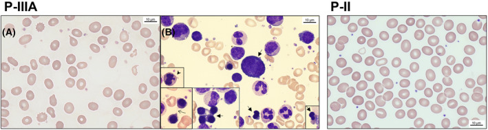
P‐IIIA: A. Peripheral blood smear had low levels of polychromasia, indicating suboptimal reticulocytosis, mild to moderate anisopoikilocytosis, normochromic macrocytes, occasional hypochromic red cells, and notable ovalocytes. B. Bone marrow aspirate showed erythroid hyperplasia with a mild left shift and rare but notable signs of dyserythropoiesis including binucleated or multinucleated erythroblasts (thick arrows), karyorrhexis (arrowhead), and even occasional cytoplasmic bridges (thin arrow). P‐II: Peripheral blood smear with low levels of polychromasia, notable anisopoikilocytosis, normochromic macrocytes, occasional hypochromic red cells, frequent ovalocytes, and several stomatocytes. [Color figure can be viewed at wileyonlinelibrary.com ]
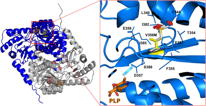
Left Panel: The image shows a surface representation overlaid on ribbon diagram of the ALAS2 protein in the active dimer (PDB ID 6HRH). Monomers are shown in blue and gray. Val356 and Y365 are shown in stick and dotted spheres representations. The pyridoxal 5′‐phosphate cofactor is shown in sphere representation. Right Panel: Val356 is shown in yellow and Met356 is shown in white; clashes are in red. The pyridoxal 5′‐phosphate is in the lower left corner (PLP). Clashes are with Leu340, Cys344, and Ile382. This figure generated with PyMOL (Schrödinger, New York, NY, USA). [Color figure can be viewed at wileyonlinelibrary.com ]
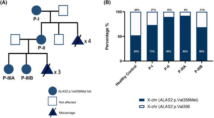
A. The family pedigree showing the affected family members reported here. Grandmother (P‐I) and mother (P‐II) suffered multiple miscarriages, likely due to embryonal lethality of males with ALAS2 Val356Met. B. X‐chromosome inactivation assay (HUMARA) results showing per cent (%) activity of the mutant (maternal) and WT (paternal) ALAS2 alleles in all affected individuals. The most severely affected members in the family (P‐II and P‐IIIA) had the maternal X‐chromosome (containing the mutant ALAS2 gene) active in at least 90% of whole‐blood genomic DNA. [Color figure can be viewed at wileyonlinelibrary.com ]
References
-
- Adam MP, Feldman J, Mirzaa GM, et al. GeneReviews. 1993.
Publication types
Grants and funding
LinkOut - more resources
Full Text Sources
Medical
