. 2024 Oct 22;98(10):e0064024.
doi: 10.1128/jvi.00640-24. Epub 2024 Sep 27.

Architectural organization and in situ fusion protein structure of lymphocytic choriomeningitis virus

Affiliations

Architectural organization and in situ fusion protein structure of lymphocytic choriomeningitis virus

Joon S Kang et al. J Virol. .

Abstract

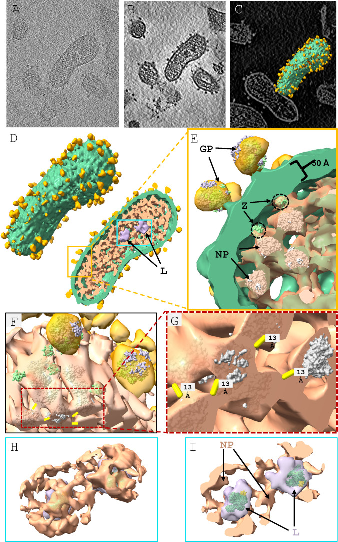
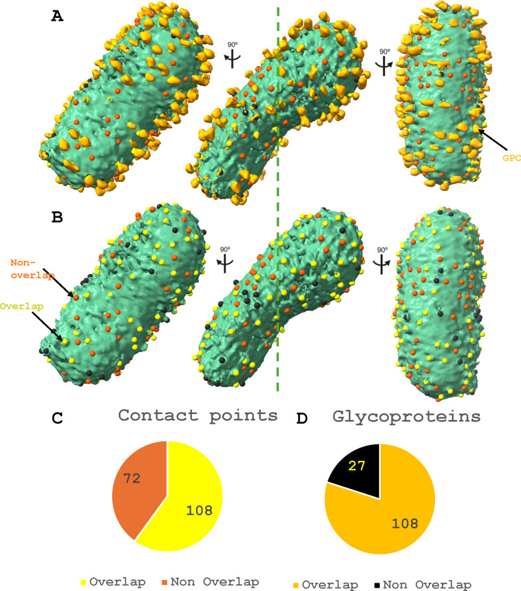
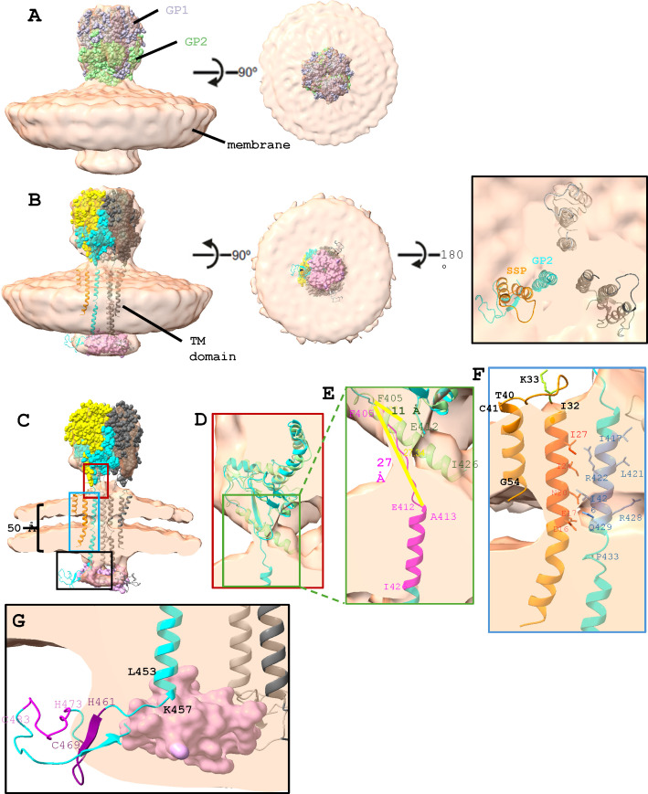
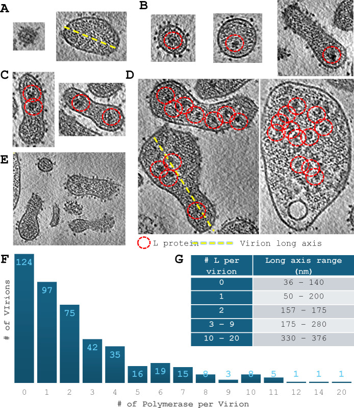
Arenaviruses exist globally and can cause hemorrhagic fever and neurological diseases, exemplified by the zoonotic pathogen lymphocytic choriomeningitis virus (LCMV). The structures of individual LCMV proteins or their fragments have been reported, but the architectural organization and the nucleocapsid assembly mechanism remain elusive. Importantly, the in situ structure of the arenavirus fusion protein complex (glycoprotein complex, GPC) as present on the virion prior to fusion, particularly with its integral stable signal peptide (SSP), has not been shown, hindering efforts such as structure-based vaccine design. Here, we have determined the in situ structure of LCMV proteins and their architectural organization in the virion by cryogenic electron tomography. The tomograms reveal the global distribution of GPC, matrix protein Z, and the contact points between the viral envelope and nucleocapsid. Subtomogram averaging yielded the in situ structure of the mature GPC with its transmembrane domain intact, revealing the GP2-SSP interface and the endodomain of GP2. The number of RNA-dependent RNA polymerase L molecules packaged within each virion varies, adding new perspectives to the infection mechanism. Together, these results delineate the structural organization of LCMV and offer new insights into its mechanism of LCMV maturation, egress, and cell entry.

Importance: The impact of COVID-19 on public health has highlighted the importance of understanding zoonotic pathogens. Lymphocytic choriomeningitis virus (LCMV) is a rodent-borne human pathogen that causes hemorrhagic fever. Herein, we describe the in situ structure of LCMV proteins and their architectural organization on the viral envelope and around the nucleocapsid. The virion structure reveals the distribution of the surface glycoprotein complex (GPC) and the contact points between the viral envelope and the underlying matrix protein, as well as the association with the nucleocapsid. The morphology and sizes of virions, as well as the number of RNA polymerase L inside each virion vary greatly, highlighting the fast-changing nature of LCMV. A comparison between the in situ GPC trimeric structure and prior ectodomain structures identifies the transmembrane and endo domains of GPC and key interactions among its subunits. The work provides new insights into LCMV assembly and informs future structure-guided vaccine design.

Keywords: arenavirus; cryogenic electron tomography; in situ structures; lymphocytic choriomeningitis virus; prefusion; spike proteins; virion.


Conflict of interest statement

The authors declare no conflict of interest.

Figures

Fig 1
Fig 1
Architectural organization of LCMV: GP-Z-NP interface and peripheral NP arrangement. (A) XY density slice view of a cryoET tomogram containing a virion of interest. (B) XY slice view of the tomogram from panel A after deconvolution and missing wedge correction. (C) Slice view of panel B with the segmented virion of interest. (D) Left: segmentation map of the virion from panel C. Right: cut-open view of the virion. (E) Zoom-in view of the yellow box region in panel D. Gold, glycoprotein; green, viral envelope; peach, nucleocapsid; and purple, L. The structures of GP (8DMI), Z (5I72), and NP (3T5N) were fitted or placed into the density as references. (F) Slightly rotated along the X-axis view of panel E with the viral envelope hidden. (G) Zoom-in view of the dotted red box region in panel F with the distance between the NPs indicated in yellow. (H and I) Segmentation of L with surrounding NP structures, where panel I is a sectional view of panel H.
Fig 2
Fig 2
Distribution of contact points and how it relates to the glycoprotein. (A and B) Center: segmentation map of a virion from Fig. 1D with the distribution of contact points between the nucleocapsid and the membrane shown as surface markers. Yellow markers indicate the contact points that coincide with the GPCs. Orange markers indicate the contact points that do not coincide with the GPCs. Black markers indicate GPCs that do not coincide with the contact points. Left and right: the view rotated 90° along the axis indicated on the center view. (B) Segmentation map of a virion from panel A with GPCs hidden to show the yellow surface markers among others, which indicate the overlap between the contact points and GPC. (C) Pie chart depicting the distribution of nucleocapsid-membrane contact points that do/do not coincide with the GPs. (D) Pie chart depicting the distribution of GPs that do/do not coincide with the nucleocapsid-membrane contact points. The color code follows the marker color code in panels A and B.
Fig 3
Fig 3
In situ structure of GPC using subtomogram averaging. (A) STA reconstruction (peach) fitted with 8DMI in space-filling model representation. GP1 is in gray, and GP2 is in light green (left, top view). (B) STA reconstruction fitted with our model and C3-symmetrized monomers of 5I72. GP1 is in yellow, GP2 is in cyan, SSP is in orange, and the Z monomer is in pink. Other GPCs are in dark gray and light gray. Ectodomain in space-filling model representation, and TM domain and C-terminal domain in cartoon representation (left, bottom view). (Right) Top view with the ectodomain removed. (C) Cut-open view of panel B. The threshold has been adjusted from panels A and B to better define the bilayer and the TM domain density. (D) Zoom-in view of the burgundy-boxed region in panel C. It shows the superimposition of 8DMI GP2 (light green) and our modeled GP2 domain (cyan). The models are in cartoon representation. (E) Zoom-in view of the green-boxed region in panel D. The region between F405 and I426 of our model is highlighted in magenta. The distance between F405 and E412 of both models is measured using the Chimera tape tool. (F) Zoom-in view of the blue-boxed region in panel C. The hydrophobic residues are labeled. SSP helix 1 residues that are potentially interacting with GP2 are highlighted in darker orange, and the GP2 residues that are interacting with SSP helix 1 and neighboring GP2 are colored in darker blue. Ectodomain and helix 2 residues of SSP are highlighted in orange. (G) Zoom-in view of the black boxed region in panel C. ZBD1 and 2 of GP2 are highlighted in dark purple and light purple, respectively. One Z monomer is displayed for clarity.
Fig 4
Fig 4
Denoised and missing-wedge corrected tomograms of LCMV virions packaged with different numbers of L. (A–E) XY slice view of denoised tomograms, highlighting virions with various sizes and numbers of polymerases. Red dashed circles indicate the polymerase complex. (A) Virions with 0 polymerase. (B) Virions with one polymerase. (C) Virions with two polymerases. (D) Virions with more than two polymerases. (E) Bird’s eye view of polymerase-packaged virions coexisting with other virions packaged with no polymerase. (F) Histogram of the number of virions versus the number of polymerases packaged in a virion. For example, there are 124 virions packaged with no polymerase. (G) Table showing the size variation of virion in relation to the number of polymerase(s) packaged in the virion. The long axis was measured as indicated in the dashed yellow line in panels A and D.

Similar articles

References

    1. Albariño CG, Palacios G, Khristova ML, Erickson BR, Carroll SA, Comer JA, Hui J, Briese T, St George K, Ksiazek TG, Lipkin WI, Nichol ST. 2010. High diversity and ancient common ancestry of lymphocytic choriomeningitis virus. Emerg Infect Dis 16:1093–1100. doi:10.3201/eid1607.091902 - DOI - PMC - PubMed
    1. Oldstone MBA. 2007. A suspenseful game of “hide and seek” between virus and host. Nat Immunol 8:325–327. doi:10.1038/ni0407-325 - DOI - PubMed
    1. Jamieson DJ, Kourtis AP, Bell M, Rasmussen SA. 2006. Lymphocytic choriomeningitis virus: an emerging obstetric pathogen? Am J Obstet Gynecol 194:1532–1536. doi:10.1016/j.ajog.2005.11.040 - DOI - PubMed
    1. Meritet JF, Krivine A, Lewin F, Poissonnier MH, Poizat R, Loget P, Rozenberg F, Lebon P. 2009. A case of congenital lymphocytic choriomeningitis virus (LCMV) infection revealed by hydrops fetalis. Prenat Diagn 29:626–627. doi:10.1002/pd.2240 - DOI - PubMed
    1. Hallam SJ, Koma T, Maruyama J, Paessler S. 2018. Review of mammarenavirus biology and replication. Front Microbiol 9:1751. doi:10.3389/fmicb.2018.01751 - DOI - PMC - PubMed

MeSH terms

Substances