Review
. 2024 Sep 22;9(9):574.
doi: 10.3390/biomimetics9090574.

Nanoparticles as Drug Delivery Vehicles for People with Cystic Fibrosis

Affiliations
Review

Nanoparticles as Drug Delivery Vehicles for People with Cystic Fibrosis

Eoin Hourihane et al. Biomimetics (Basel). .

Abstract

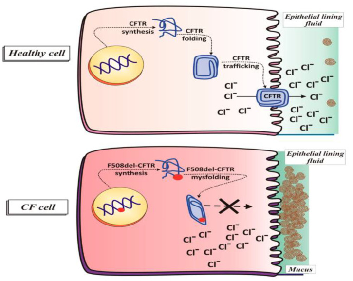
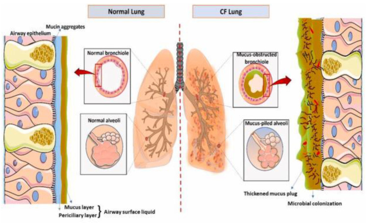
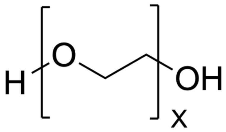
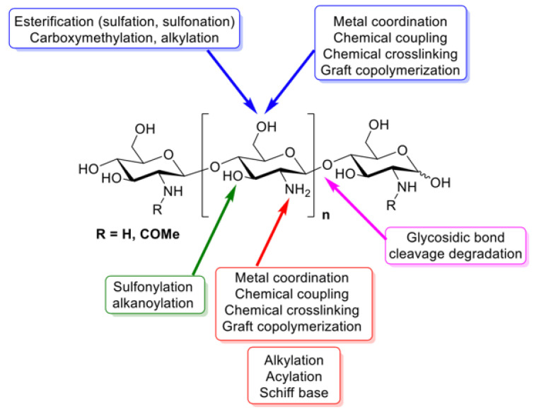
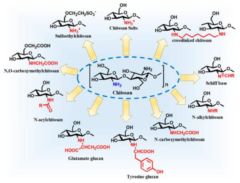
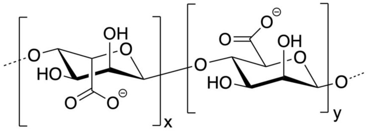
Cystic Fibrosis (CF) is a life-shortening, genetic disease that affects approximately 145,000 people worldwide. CF causes a dehydrated mucus layer in the lungs, leading to damaging infection and inflammation that eventually result in death. Nanoparticles (NPs), drug delivery vehicles intended for inhalation, have become a recent source of interest for treating CF and CF-related conditions, and many formulations have been created thus far. This paper is intended to provide an overview of CF and the effect it has on the lungs, the barriers in using NP drug delivery vehicles for treatment, and three common material class choices for these NP formulations: metals, polymers, and lipids. The materials to be discussed include gold, silver, and iron oxide metallic NPs; polyethylene glycol, chitosan, poly lactic-co-glycolic acid, and alginate polymeric NPs; and lipid-based NPs. The novelty of this review comes from a less specific focus on nanoparticle examples, with the focus instead being on the general theory behind material function, why or how a material might be used, and how it may be preferable to other materials used in treating CF. Finally, this paper ends with a short discussion of the two FDA-approved NPs for treatment of CF-related conditions and a recommendation for the future usage of NPs in people with Cystic Fibrosis (pwCF).

Keywords: FDA; antibiotics; cystic fibrosis; drug delivery; lipids; metals; nanoparticles; polymers; treatments.


Conflict of interest statement

The authors declare no conflicts of interest.

Figures

Figure 1
Figure 1
CFTR function in healthy and diseased cells. CFTR transports Cl and HCO3 from inside the cell to outside the cell in epithelial cells in the lungs. This movement of anions forces Na+ and water to move in the same direction as Cl and HCO3. This water is then used to hydrate the mucosal layer of the lungs. The lack of a functional CFTR protein causes the mucosal layer to dehydrate and shrink. Not shown in this figure is the movement of water, sodium, and bicarbonate [10]. Reprinted from “An Intriguing Involvement of Mitochondria in Cystic Fibrosis,” by M. Favia, L. de Bari, A. Bobba, and A. Atlante, 2019, Journal of Clinical Medicine, 8(11), p. 1890 (https://doi.org/10.3390/jcm8111890). CC BY-NC.
Figure 2
Figure 2
Normal lung vs. CF lung [23]. Reprinted from “Role of Innate Immunity and Systemic Inflammation in Cystic Fibrosis Disease Progression” by A.K. Purushothaman and E.J.R. Nelson, 2023, Heliyon, 9(7), p. 5 (https://doi.org/10.1016/j.heliyon.2023.e17553). CC BY-NC.
Figure 3
Figure 3
Deposition of particles in the lungs based on size. Inhaled particles deposit in the lungs primarily based on size. NPs 5–10 μm will only make it as far as the nasal cavity. NPs 3–5 μm will make it as far as the trachea, NPs 2–3 μm as far as the bronchus, and NPs 1–2 μm as far as the bronchiole. NPs 1–1000 nm will make it as far as the alveoli, though they will deposit everywhere in the lungs [29]. Reprinted from “Exposure to Inorganic Nanoparticles: Routes of Entry, Immune Response, Biodistribution and In Vitro/In Vivo Toxicity Evaluation,” by V. De Matteis, 2017, Toxics, 5(4), p. 29 (https://doi.org/10.3390/toxics5040029). CC BY-NC.
Figure 4
Figure 4
Ferromagnetic particles and SPIONs. Change in magnetic moments of ferromagnetic particles versus superparamagnetic particles. (A) In the absence of a magnetic field, ferromagnetic particles have magnetic moments in many directions, often aligned with their grain, while SPIONs have a single magnetic moment. (B) In the presence of a magnetic field, ferromagnetic particles and SPIONs align their magnetic moments to be parallel with the external magnetic field. (C) Upon removal of the external magnetic fields, ferromagnetic particles retain some of the magnetization as they dissipate energy and slowly return to the state present in A. SPIONs immediately release their magnetization and return back to the state present in A [48]. Reprinted from “Chapter 11—Magnetic Nanoparticles for Application in Biomedical Sensing”, by D. Alcantara and L. Josephson, 2012, Frontiers of Nanoscience, Vol. 4, p. 270 (https://doi.org/10.1016/B978-0-12-415769-9.00011-X). Copyright 2012 by Elsevier. Reprinted with permission.
Figure 5
Figure 5
PEG structure [64].
Figure 6
Figure 6
Groups in chitosan that can be functionalized [70]. Reprinted from “Chitosan: An Overview of Its Properties and Applications,” by I. Aranaz, A. Alcántara, M.C. Civera, C. Arias, B. Elorza, A.H. Caballero, and N. Acosta, 2021, Polymers, 13(19), p. 3256 (https://doi.org/10.3390/polym13193256). CC BY-NC.
Figure 7
Figure 7
Chitosan derivatives [69]. Reprinted from “Chitosan Nanoparticles-Insight into Properties, Functionalization and Applications in Drug Delivery and Theranostics,” by J. Jhaveri, Z. Raichura, T. Khan, M. Momin, and A. Omri, 2021, Molecules, 26(2), p. 272 (https://doi.org/10.3390/molecules26020272). CC BY-NC.
Figure 8
Figure 8
PLGA structure. Lactic acid monomers on left (x) and glycolic acid monomers on right (y) [64].
Figure 9
Figure 9
Alginate structure. Sodium ions coordinate around COO groups in sugars. G on left, M on right [64].
Figure 10
Figure 10
Alginate egg-box model (a) Ionic crosslinking as depicted on the molecular level. (b) Schematic view of crosslinking with full crosslinking across all alginate molecules. (c) Schematic view of crosslinking with partially crosslinked alginate molecules [87]. Adapted from “Ion-Induced Polysaccharide Gelation: Peculiarities of Alginate Egg-Box Association with Different Divalent Cations,” by A. Makarova, S. Derkach, T. Khair, M. Kazantseva, Y. Zuev, and O. Zueva, 2023, Polymers, 15(5), p. 1243 (https://doi.org/10.3390/polym15051243). CC BY-NC.
Figure 11
Figure 11
Types of lipid-based NPs. (a) Liposomes are formed by phospholipids and may encapsulate drugs in the hydrophilic core or hydrophobic section of the tails. (b) Solid Lipid Nanoparticles are micelles formed with a surface active agent (typically a phospholipid) along with a solid lipid. Hydrophobic drugs may be encapsulated in the solid lipid phase, or a smaller micelle may form inside the particle to encapsulate hydrophilic drugs. (c) Nanostructured lipid carries are micelles formed with a surface active agent (typically a phospholipid) along with both solid and liquid phase lipids. Hydrophobic drugs are encapsulated within the solid and liquid phase lipid. (d) Hybrid lipid-polymeric NPs are micelles formed by encapsulating a polymeric NP with phospholipid. Hydrophobic drugs are encapsulated by the polymer [102]. Reprinted from “Lipid Nanoparticles for Drug Delivery,” by L. Xu, X. Wang, Y. Liu, G. Yang, R. Falconer, and C. Zhao, 2021, Advanced NanoBiomed Research, 2(2) (https://doi.org/10.1002/anbr.202100109). CC BY-NC.
Figure 12
Figure 12
Phospholipid polar head group modifications [104]. Reprinted from “Liposome surface modification by phospholipid chemical reactions,” by P. de Lima, A. Butera, L. Cabeça, and R. Ribeiro-Viana, 2021, Chemistry and Physics of Lipids, 237 (https://doi.org/10.1016/j.chemphyslip.2021.105084). Copyright 2021 by Elsevier. Reprinted with permission.
Figure 13
Figure 13
Lipid-NP functionalization [106]. Reprinted from “Liposomes or Extracellular Vesicles: A Comprehensive Comparison of Both Lipid Bilayer Vesicles for Pulmonary Drug Delivery,” by A. Al-Jipouri, S. Almurisi, K. Al-Japairai, L. Bakar, and A. Doolaanea, 2023, Polymers, 15(2), p. 318 (https://doi.org/10.3390/polym15020318). CC BY-NC.

Similar articles

References

    1. Lyczak J.B., Cannon C.L., Pier G.B. Lung Infections Associated with Cystic Fibrosis. Clin. Microbiol. Rev. 2002;15:194–222. doi: 10.1128/CMR.15.2.194-222.2002. - DOI - PMC - PubMed
    1. About Cystic Fibrosis | Cystic Fibrosis Foundation. [(accessed on 21 April 2024)]. Available online: https://www.cff.org/intro-cf/about-cystic-fibrosis.
    1. McBennett K.A., Davis P.B., Konstan M.W. Increasing Life Expectancy in Cystic Fibrosis: Advances and Challenges. Pediatr. Pulmonol. 2022;57:S5–S12. doi: 10.1002/ppul.25733. - DOI - PMC - PubMed
    1. Understanding Changes in Life Expectancy | Cystic Fibrosis Foundation. [(accessed on 21 April 2024)]. Available online: https://www.cff.org/managing-cf/understanding-changes-life-expectancy.
    1. Harris A. Cystic Fibrosis Gene. Br. Med. Bull. 1992;48:738–753. doi: 10.1093/oxfordjournals.bmb.a072575. - DOI - PubMed

LinkOut - more resources