Review
. 2024 Sep 12;11(9):910.
doi: 10.3390/bioengineering11090910.

An Insert Goniometer Can Help Select the Optimal Insert Thickness When Performing Kinematically Aligned Total Knee Arthroplasty with a Medial 1:1 Ball-in-Socket and Lateral Flat Surface Insert and Posterior Cruciate Ligament Retention

Affiliations
Review

An Insert Goniometer Can Help Select the Optimal Insert Thickness When Performing Kinematically Aligned Total Knee Arthroplasty with a Medial 1:1 Ball-in-Socket and Lateral Flat Surface Insert and Posterior Cruciate Ligament Retention

Sahil A Sanghavi et al. Bioengineering (Basel). .

Abstract

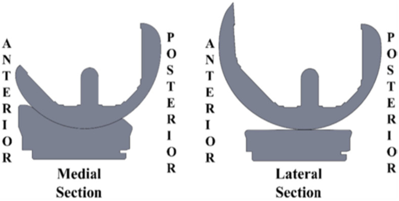
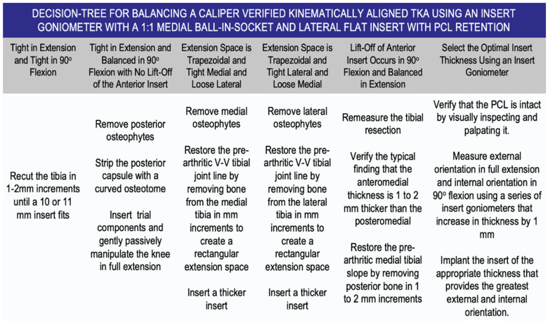
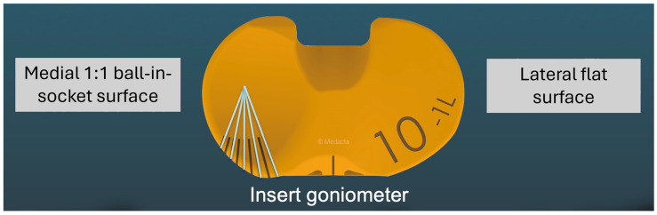
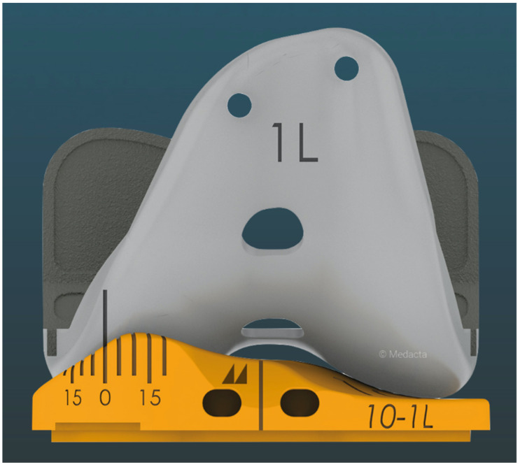
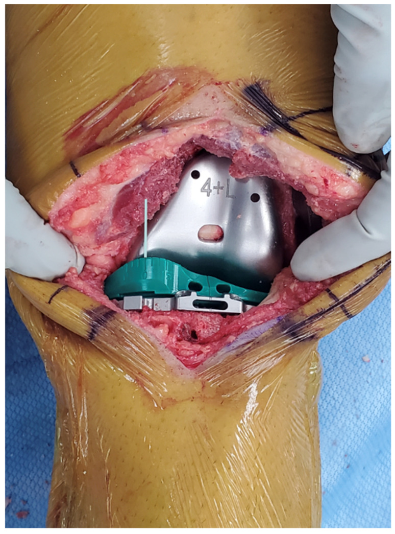
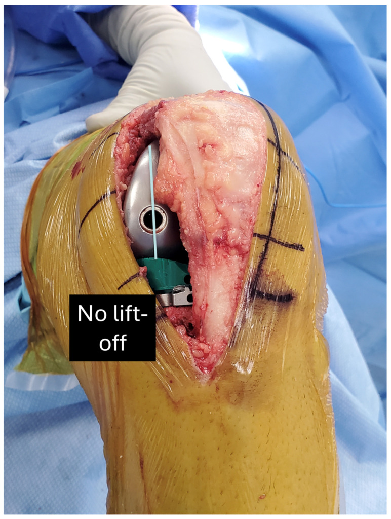
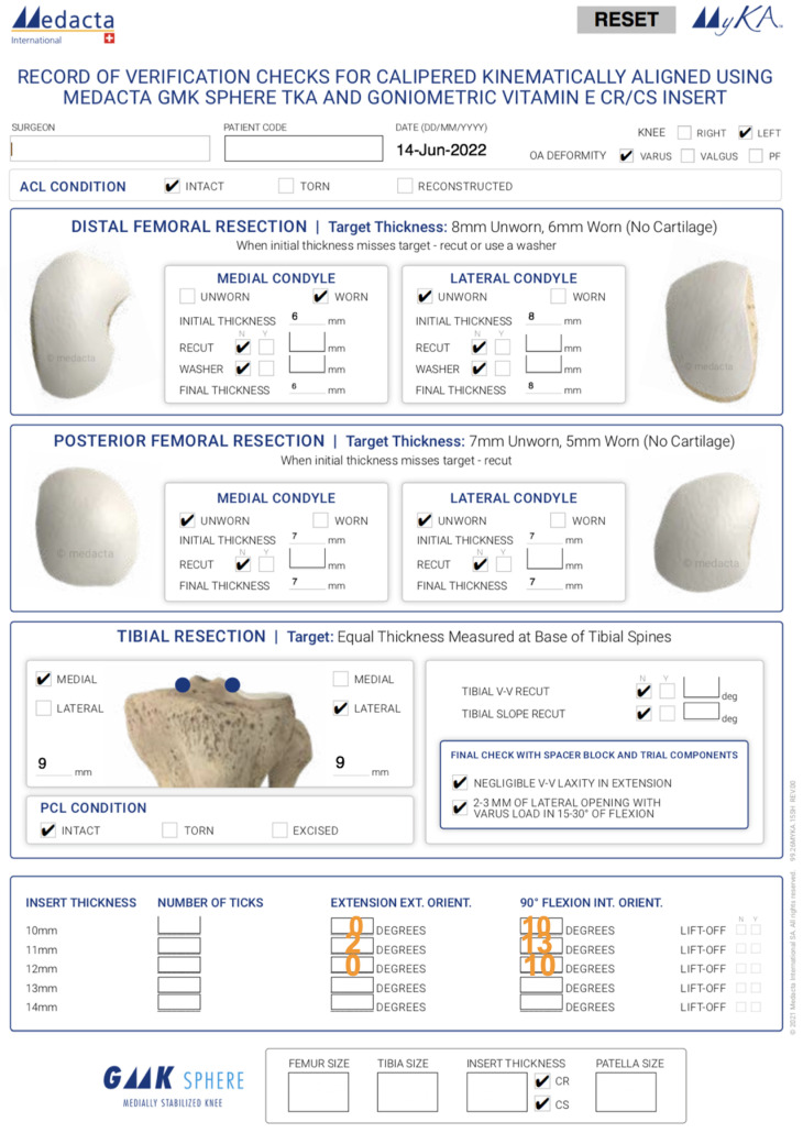
Current surgical practices in total knee arthroplasty (TKA) have advanced and include significant changes and improvements in alignment philosophies, femorotibial implant conformities, and ligament management to replicate in vivo knee kinematics. While corrective measures have emphasized sagittal plane alignment to restore normal flexion-extension (F-E) motion and coronal plane ligament balance, internal-external (I-E) rotation kinematics in the axial plane have been largely neglected. Recent in vivo evidence indicates that the combination of factors necessary to closely restore native tibial rotation as the knee flexes and extends is kinematic alignment (KA), which resurfaces the patient's pre-arthritic knee without releasing ligaments, an insert with medial 1:1 ball-in-socket conformity and a lateral flat surface, and posterior cruciate ligament (PCL) retention. However, the inherent anterior-posterior (A-P) stability provided by the medial 1:1 ball-in-socket limits the surgeon's ability to select the correct insert thickness using manual laxity testing. Accordingly, this review presents the design and validation of an instrument called an insert goniometer that measures I-E tibial rotation for inserts that differ in thickness by 1 mm and uses rotation limits at extension and 90° flexion to select the optimal insert thickness. The optimal thickness is the one that provides the greatest external tibial orientation in extension and internal tibial orientation at 90° flexion without lift-off of the insert.

Keywords: external tibial rotation; internal tibial rotation; quadriceps line of force; screw home mechanism; tibiofemoral kinematics; total knee replacement.


Conflict of interest statement

S.M.H. is a paid consultant, a paid speaker, and receives royalties from Medacta International. A.J.N. is a paid consultant and receives royalties from Medacta International. M.L.H. receives research support from Medacta USA, Inc. and is on the editorial board of the Journal of Biomechanics.

Figures

Figure 1
Figure 1
Section views of the femoral condyles and tibial insert in the A–P direction and through the dwell point of the medial 1:1 ball-in-socket insert showing articular geometry in the medial and lateral compartments.
Figure 2
Figure 2
Composite showing the decision tree followed by the surgeon when he/she performs caliper-verified KA TKA. The technique sets the components to restore the pre-arthritic distal and posterior femoral and proximal tibial joint lines within 0 ± 0.5 mm, which restores native tibial compartment forces without the release of healthy ligaments, including the PCL.
Figure 3
Figure 3
Image showing the design of the insert goniometer, which consists of lines in 5° increments that are calibrated to the axis of rotation or pivot point of the knee in the medial compartment. The lines extend over the anterior surface (see Figure 4) so that the I–E orientation of the tibia relative to the femur can be indicated.
Figure 4
Figure 4
Image showing the method used by the insert goniometer to measure I–E orientation of the tibia relative to the femur. A reference line is laser etched vertically on the center of the medial condyle of the trial femoral component. As the tibia rotates internally/externally during flexion/extension, respectively, the I–E orientation is indicated. In this figure where the knee is extended, the indicated external orientation is 0°.
Figure 5
Figure 5
Photo of the insert goniometer at extension for an example case. The 10 mm thick insert goniometer had an external tibial orientation that measured 0°.
Figure 6
Figure 6
Photo of the insert goniometer at 90° flexion for an example case. There was no lift-off of the insert goniometer. The internal tibial orientation with the 10 mm thick insert goniometer was 10°, and there was no liftoff.
Figure 7
Figure 7
Verification check sheet for a caliper-verified KA TKA, which documents the thickness of the bony resections and the internal–external tibial orientations mentioned in the example case.

Similar articles

References

    1. French S.R., Munir S., Brighton R. A single surgeon series comparing the outcomes of a cruciate retaining and medially stabilized total knee arthroplasty using kinematic alignment principles. J. Arthroplast. 2020;35:422–428. doi: 10.1016/j.arth.2019.09.021. - DOI - PubMed
    1. Scott D.F., Gray C.G. Outcomes are better with a medial-stabilized vs a posterior-stabilized total knee implanted with kinematic alignment. J. Arthroplast. 2022;37:S852–S858. doi: 10.1016/j.arth.2022.02.059. - DOI - PubMed
    1. Griffin F.M., Insall J.N., Scuderi G.R. Accuracy of soft tissue balancing in total knee arthroplasty. J. Arthroplast. 2000;15:970–973. doi: 10.1054/arth.2000.6503. - DOI - PubMed
    1. Gu Y., Howell S.M., Hull M.L. Simulation of total knee arthroplasty in 5 degrees or 7 degrees valgus: A study of gap imbalances and changes in limb and knee alignments from native. J. Orthop. Res. 2017;35:2031–2039. doi: 10.1002/jor.23492. - DOI - PubMed
    1. Heesterbeek P.J., Wymenga A.B. PCL balancing, an example of the need to couple detailed biomechanical parameters with clinical functional outcome. Knee Surg. Sports Traumatol. Arthrosc. 2010;18:1301–1303. doi: 10.1007/s00167-010-1254-6. - DOI - PubMed