Review
. 2024 Sep 12;11(9):912.
doi: 10.3390/bioengineering11090912.

Optical Image Sensors for Smart Analytical Chemiluminescence Biosensors

Affiliations
Review

Optical Image Sensors for Smart Analytical Chemiluminescence Biosensors

Reza Abbasi et al. Bioengineering (Basel). .

Abstract

Optical biosensors have emerged as a powerful tool in analytical biochemistry, offering high sensitivity and specificity in the detection of various biomolecules. This article explores the advancements in the integration of optical biosensors with microfluidic technologies, creating lab-on-a-chip (LOC) platforms that enable rapid, efficient, and miniaturized analysis at the point of need. These LOC platforms leverage optical phenomena such as chemiluminescence and electrochemiluminescence to achieve real-time detection and quantification of analytes, making them ideal for applications in medical diagnostics, environmental monitoring, and food safety. Various optical detectors used for detecting chemiluminescence are reviewed, including single-point detectors such as photomultiplier tubes (PMT) and avalanche photodiodes (APD), and pixelated detectors such as charge-coupled devices (CCD) and complementary metal-oxide-semiconductor (CMOS) sensors. A significant advancement discussed in this review is the integration of optical biosensors with pixelated image sensors, particularly CMOS image sensors. These sensors provide numerous advantages over traditional single-point detectors, including high-resolution imaging, spatially resolved measurements, and the ability to simultaneously detect multiple analytes. Their compact size, low power consumption, and cost-effectiveness further enhance their suitability for portable and point-of-care diagnostic devices. In the future, the integration of machine learning algorithms with these technologies promises to enhance data analysis and interpretation, driving the development of more sophisticated, efficient, and accessible diagnostic tools for diverse applications.

Keywords: biosensor; chemiluminescence; image sensors; machine learning; optical.


Conflict of interest statement

R.A., X.H., and S.W.-H. are co-founders of a 2023 McGill spinoff company, Phoela Health Inc.

Figures
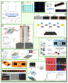
Figure 3
Figure 3
Electrochemiluminescence with single-electrode (SE) configuration: (a) schematic of SE ECL on CMOS image sensor (top), structure of SE configuration, and distribution of potential along the single electrode (bottom). Adapted with permission from [104], Copyright (2024). (b) First demonstration of SE-ECL, multiplex ECL using SE configuration. Adapted with permission from [106], Copyright (2018). SE-ECL for single and multiplex experiments are created by attaching labels with varying hole patterns onto ITO electrodes. (c) Paper-based SE-ECL on carbon ink. Adapted with permission from [146], Copyright (2022). A paper-based biosensor used glow sticks as a luminophore, detected by a smartphone. A two-compartment paper device enabled separate optimization of sensing and detection reactions. (d) Fabrication of multiple-well SE-ECL on carbon ink (top) and equivalent circuit (bottom) for immunoassay application. Adapted with permission from [147], Copyright (2023). This paper-based SE-ECL, featuring a perforated sticker on a carbon ink electrode, utilized antibody immobilization and Co–Pt nanoparticles for sensitive SARS-CoV-2 detection. (e) SE-ECL with multiple channels fabricated by punching polypyrrole film and attaching it to the carbon ink layer (top) and ECL reactions on the cathodic and anodic side of each well (bottom). Adapted with permission from [148], Copyright (2023). (f) Paper-based SE-ECL fabricated by reducing graphene oxide via laser and wax printing to form microfluidic channels for glucose and lactate sensing. Adapted with permission from [149], Copyright (2024). (g) Multiplex SE-ECL fabricated using Ru(phen)32+ modified carbon nanotube/graphene film and a plastic sticker with 24 holes for dopamine detection. Adapted with permission from [150], Copyright (2024). (h) SE-ECL device fabricated using laser-induced graphene and polyimide film. Adapted with permission from [151], Copyright (2024). (i) First multicolor SE-ECL device fabricated by 3D-printed channels. Multicolor ECL using different potential in different channels (top), multicolor ECL using potential gradient along the SE (bottom left), photo of the 3D-printed SE-ECL (bottom center), and multicolor SE-ECL on different channels (bottom right). Adapted with permission from [105], Copyright (2024). (j) Miniaturized and microfluidic-integrated SE-ECL on CMOS image sensor: schematic of the device (left) and detection of multiple analytes including glucose and uric acid with this device (right). Adapted with permission from [105], Copyright (2024).
Figure 1
Figure 1
Overview of the analytical chemiluminescence sensors: (a) application of analytical chemiluminescence in food safety, health monitoring, and environmental monitoring; (b) optical phenomena including bioluminescence, chemiluminescence, and electrochemiluminescence; (c) optical detectors from point detectors (left) including photomultiplier tub and avalanche photodiode to multiplexing (plate reader) (d) optical image sensors including CCD and CMOS image sensors.
Figure 2
Figure 2
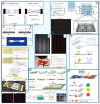
Electrochemiluminescence with bipolar configuration: (a) Electrode configuration in conventional ECL with a three-electrode setup (left), and in bipolar ECL (BPE) where a conductor is placed between the electrochemical cell, and the potential difference across the conductor drives the ECL reaction (right). Adapted with permission from [135], Copyright (2024). (b) Bipolar electrode array for multiplexed detection of prostate cancer biomarkers. Adapted with permission from [136], Copyright (2022). Liquid flow is directed by using a specialized channel structure that leverages differential flow resistance (c) paper-based BPE ECL for imaging and sensing. Adapted with permission from [137], Copyright (2024). Microfluidic channels are fabricated on filter paper using wax screen-printing, while carbon ink-based BPE and driving electrodes are also screen-printed onto the paper. (d) ECL using 1000 BPE arrays. Optical image (top left), ECL image (top right), electrochemical cell setup (bottom left), and ECL intensity profile (bottom right) of these arrays. Adapted with permission from [138], Copyright (2010). The simplified device uses a single pair of driving electrodes to activate a 500 μm × 50 μm electrode array, and the BPEs are placed in a shallow electrolyte pool between parallel plates to ensure uniform electric field (e) porous BPE using ITO nanoparticles. Top view and cross-section (top) and H2O2 sensing (bottom). Adapted with permission from [139], Copyright (2024). PDMS cover with 90 μm microchannels seals the BPE channel, separating detection and reporting channels. A nanoporous ITO layer detects H2O2, while a bare ITO layer reports ECL. The PDMS cover and ITO-coated glass form the complete BPE microchip. (f) BPE arrays for ECL imaging and multiplex sensing. Schematic of the device (top), real device, and ECL images (bottom). Adapted with permission from [140], Copyright (2022). The device features integrated cathodic and anodic poles and leads, insulated by a polyimide layer. Platinum and gold BPEs were fabricated using arrays of these poles. (g) Fabrication process of paper-based PBE aptasensor for detection of carcinoembryonic antigen. Adapted with permission from [141], Copyright (2024). Janus-like gold-coated Fe3O4 nanospheres were synthesized in this device and showed catalytic activity for H2O2 reduction. (h) Multicolor ECL using BPE configuration to detect prostate-specific antigen. Adapted with permission from [142], Copyright (2024). Selective ECL excitation is enabled by tuning the interfacial potential of the BPE poles in this device. (i) BPE ECL sensor using gold and glassy carbon beads for detection of tetracycline. Adapted with permission from [143], Copyright (2017). Au particles were selectively deposited on one side of a GCB electrode, controlling coverage from 0 to 45.3% using bipolar electroplating. This modified GCB allows for varying biomolecule conjugation.

Similar articles

Cited by

References

    1. de Araujo W.R., Lukas H., Torres M.D.T., Gao W., de la Fuente-Nunez C. Low-Cost Biosensor Technologies for Rapid Detection of COVID-19 and Future Pandemics. ACS Nano. 2024;18:1757–1777. doi: 10.1021/acsnano.3c01629. - DOI - PMC - PubMed
    1. Jiménez-Rodríguez M.G., Silva-Lance F., Parra-Arroyo L., Medina-Salazar D.A., Martínez-Ruiz M., Melchor-Martínez E.M., Martínez-Prado M.A., Iqbal H.M.N., Parra-Saldívar R., Barceló D., et al. Biosensors for the detection of disease outbreaks through wastewater-based epidemiology. Trends Anal. Chem. 2022;155:116585. doi: 10.1016/j.trac.2022.116585. - DOI - PMC - PubMed
    1. Cheon J., Qin J., Lee L.P., Lee H. Advances in Biosensor Technologies for Infection Diagnostics. Acc. Chem. Res. 2022;55:121–122. doi: 10.1021/acs.accounts.1c00647. - DOI - PubMed
    1. Teles F.S.R.R., de Távora Tavira L.A.P., da Fonseca L.J.P. Biosensors as rapid diagnostic tests for tropical diseases. Crit. Rev. Clin. Lab. Sci. 2010;47:139–169. doi: 10.3109/10408363.2010.518405. - DOI - PubMed
    1. Lin P.-H., Sheu S.-C., Chen C.-W., Huang S.-C., Li B.-R. Wearable hydrogel patch with noninvasive, electrochemical glucose sensor for natural sweat detection. Talanta. 2022;241:123187. doi: 10.1016/j.talanta.2021.123187. - DOI - PubMed

LinkOut - more resources