. 2024 Sep 21;11(9):946.
doi: 10.3390/bioengineering11090946.

A 3D Epithelial-Mesenchymal Co-Culture Model of the Airway Wall Using Native Lung Extracellular Matrix

Affiliations

A 3D Epithelial-Mesenchymal Co-Culture Model of the Airway Wall Using Native Lung Extracellular Matrix

Roderick H J de Hilster et al. Bioengineering (Basel). .

Abstract

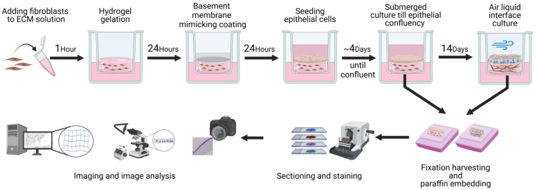
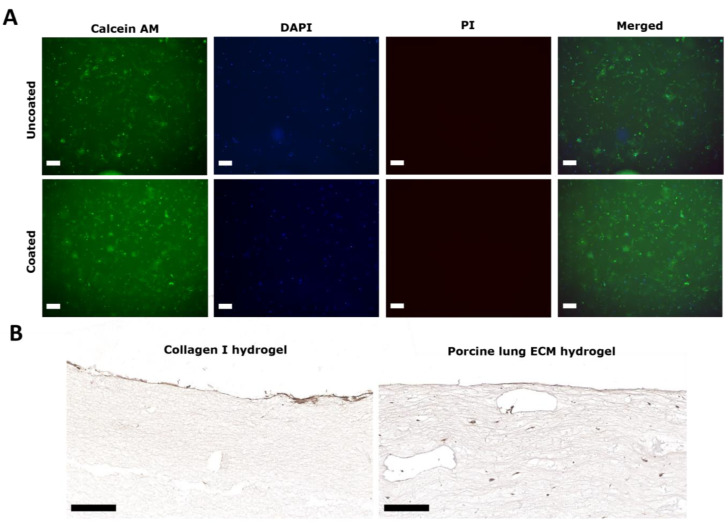
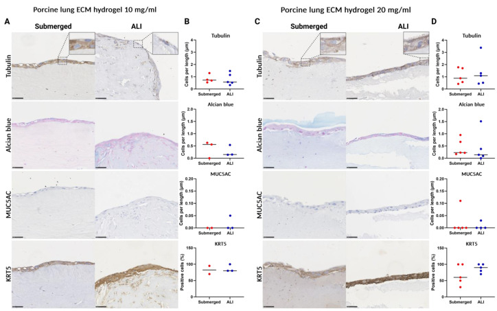
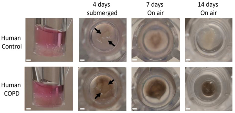
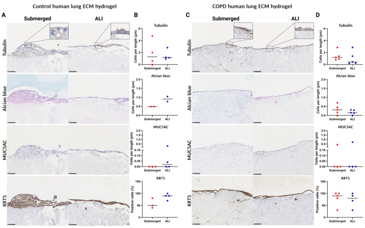
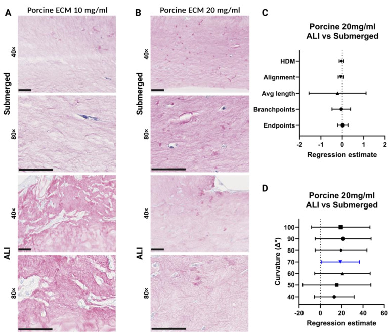
Chronic obstructive pulmonary disease (COPD) is a chronic lung disease characterized by ongoing inflammation, impaired tissue repair, and aberrant interplay between airway epithelium and fibroblasts, resulting in an altered extracellular matrix (ECM) composition. The ECM is the three-dimensional (3D) scaffold that provides mechanical support and biochemical signals to cells, now recognized not only as a consequence but as a potential driver of disease progression. To elucidate how the ECM influences pathophysiological changes occurring in COPD, in vitro models are needed that incorporate the ECM. ECM hydrogels are a novel experimental tool for incorporating the ECM in experimental setups. We developed an airway wall model by combining lung-derived ECM hydrogels with a co-culture of primary human fibroblasts and epithelial cells at an air-liquid interface. Collagen IV and a mixture of collagen I, fibronectin, and bovine serum albumin were used as basement membrane-mimicking coatings. The model was initially assembled using porcine lung-derived ECM hydrogels and subsequently with COPD and non-COPD human lung-derived ECM hydrogels. The resulting 3D construct exhibited considerable contraction and supported co-culture, resulting in a differentiated epithelial layer. This multi-component 3D model allows the investigation of remodelling mechanisms, exploring ECM involvement in cellular crosstalk, and holds promise as a model for drug discovery studies exploring ECM involvement in cellular interactions.

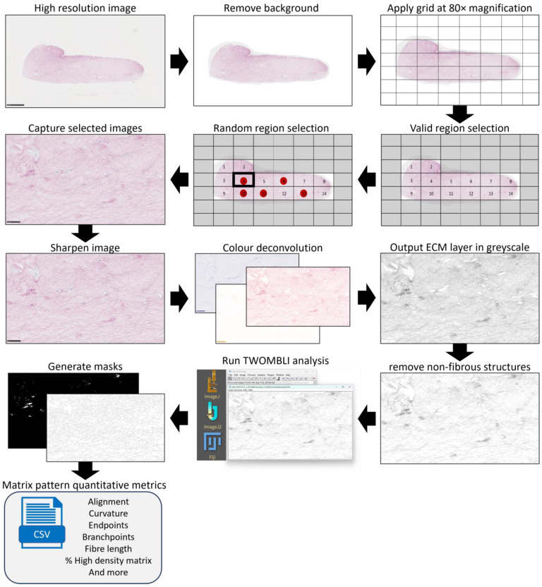
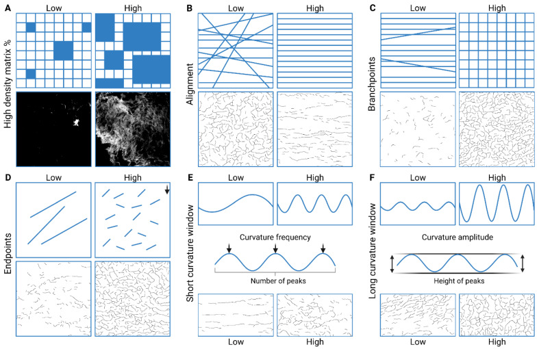
Keywords: COPD; TWOMBLI; extracellular matrix.


Conflict of interest statement

The authors declare no conflicts of interest. The funders had no role in the design of the study; in the collection, analyses, or interpretation of data; in the writing of the ma script, or in the decision to publish the results.

Figures

Figure 1
Figure 1
Schematic representation of the model assembly steps. Workflow for the preparation and analysis of the 3D co-culture of primary human fibroblasts and epithelial cells with lung ECM hydrogels. Created with biorender.com.
Figure 2
Figure 2
TWOMBLI analysis image preparation. Workflow for the preparation of Picrosirius red-stained (PSR) high-resolution images for TWOMBLI analysis of extracellular matrix organization. Eligible regions (those containing ECM hydrogel with no major defects and abnormalities) were identified, and then, 5 regions of interest were selected through a random number generator. Images were taken at 80× magnification of the selected regions and sharpened. These images were then processed using Fiji ImageJ to separate the specific signal for the red (PSR) signal. Lastly, the TWOMBLI macro generated high-density matrix and fibre masks from the processed images, from which the quantitative metrics were generated.
Figure 3
Figure 3
TWOMBLI analysis matrix metrics. Images show schematic examples of matrix metrics together with examples of TWOMBLI-generated masks from the Picrosirius red-stained ECM hydrogels. (A) Percentage of high-density matrix indicating accumulated matrix. (B) Alignment of matrix fibres. (C) Branchpoints: the number of intersections of mask fibres in the image. (D) Endpoints: number of fibre end points. (E) Short-curvature windows: describing the frequency of the peaks in the fibre curvature. (F) Long-curvature windows: describing the amplitude of the peaks in the fibre curvature. Adapted from [57] (published under CC-BY 4.0). Created with BioRender.com.
Figure 4
Figure 4
Collagen IV coating of collagen I and porcine lung ECM hydrogels on cell viability. (A) Collagen I-embedded fibroblast viability 1 day after collagen IV staining procedure. Calcein AM (green) staining of live fibroblasts; propidium iodine (PI; red) staining of dead cells; DAPI (blue) indicating nuclei; and a brightfield overview. The scale bars represent 200 μm. (B) Immunohistochemical staining of collagen IV on top of fibroblast-seeded collagen I and 10 mg/mL porcine lung ECM hydrogels 24 h after coating. The scale bars represent 100 μm. Results are representative for all experiments (n = 2).
Figure 5
Figure 5
Porcine lung ECM hydrogel’s Young’s modulus. Comparison of stiffness of porcine lung ECM hydrogels prepared either at a concentration of 10 mg/mL or 20 mg/mL. For each hydrogel condition, 3 replicate gels were made and measured individually on 2 separate occasions. Mann–Whitney U test was used to compare the two concentrations; * p < 0.005.
Figure 6
Figure 6
Model assembly with porcine lung ECM hydrogel and its contraction over time. (A) Macroscopic view of the 10 and 20 mg/mL porcine lung ECM hydrogel 3D co-culture model: side view directly after full assembly; top view after 4 days submerged culture; 7-day ALI culture; and 14-day ALI culture. (B) Microscopic (20×) images after initial seeding of epithelial cells on and fibroblast seeding in ECM hydrogel showing reduced visibility after hydrogel contraction. (C) Brightfield and fluorescent microscopic images (10×): images 1 and 5 days after epithelial cell seeding on top of fibroblast-containing or empty 10 mg/mL porcine lung ECM hydrogels. Cells are stained with DAPI to visualize nuclei. ECM: extracellular matrix, ALI: air–liquid interface, FN: fibronectin, BSA: bovine serum albumin, DAPI: 4′,6-diamidino-2-phenylindole.
Figure 7
Figure 7
Different porcine lung ECM hydrogel contraction outcomes. Haematoxylin and eosin-stained porcine lung ECM hydrogel co-culture model after 14 days of air–liquid interface culture. (A) Epithelial confluency spanning from the hydrogel across the membrane. (B) Epithelial confluence covering the entire contracted ECM and small enveloped accumulated cell pockets (arrow). (C) Folding of the entire hydrogel during contraction. Scale bars are 500 µm. ECM: extracellular matrix.
Figure 8
Figure 8
Histological analysis of primary airway epithelial cell differentiation prior to and after air–liquid interface (ALI) culture. Tubulin immunostaining (ciliated cells), Alcian blue staining (goblet cells), MUC5AC immunostaining (club cells), and KRT5 immunostaining (basal cells). Submerged culture was fixed after epithelial cells reached confluence, whereas ALI culture exposed the submerged cultured cells to air for 14 days before fixation. (A) Intact control human lung. (B) Representative images of sections (40× magnification) of submerged- and ALI-cultured airway epithelial cells on top of a collagen I–fibronectin–bovine serum albumin-coated membrane. (C). Quantified differentiated epithelial cells on membranes coated with collagen I-FN-BSA. (D). Representative images of sections (40× magnification) of submerged- and ALI-cultured airway epithelial cells on top of a collagen IV-coated membrane. (E) Quantified differentiated epithelial cells on membranes coated with collagen IV. Scale bars are 50 µm. All data are shown as means with all available data plotted individually (n = 5). FN: fibronectin, BSA: bovine serum albumin, ALI: air–liquid interface, MUC5AC: mucin-5AC, KRT5: keratin 5. Made with Biorender.com.
Figure 9
Figure 9
Histological analysis of primary airway epithelial, fibroblast, and porcine lung ECM co-culture model of cell differentiation prior to and after air–liquid interface (ALI) culture. Tubulin immunostaining (ciliated cells), Alcian blue staining (goblet cells), MUC5AC immunostaining (club cells), and KRT5 immunostaining (basal cells). Submerged culture was fixed after epithelial cells reached confluence, whereas ALI culture exposed the submerged cultured cells to air for 14 days before fixation. (A) Representative images of sections (40× magnification) of submerged- and ALI-cultured porcine ECM (conc. 10 mg/mL) epithelial cell fibroblast co-culture model. (B). Quantified differentiated epithelial cells on membranes coated with collagen I-FN-BSA. (C) Representative images of sections (40× magnification) of porcine ECM (conc. 20 mg/mL) epithelial cell fibroblast co-culture model. (D) Quantified differentiated epithelial cells on membranes coated with collagen IV. Scale bars are 50 µm. All data are shown as means with all available data plotted individually (n = 1–5). ECM: extracellular matrix, ALI: air–liquid interface, MUC5AC: mucin-5AC, KRT5: keratin 5. Made with Biorender.com.
Figure 10
Figure 10
Model assembly with human lung ECM hydrogel and its contraction over time. Macroscopic view of the control and COPD human lung-derived ECM hydrogel 3D co-culture model: side view directly after full assembly; top view after 4 days submerged culture; 7-day ALI culture; and 14-day ALI culture.
Figure 11
Figure 11
Histological analysis of primary airway epithelial, fibroblast, and human lung ECM (HECM) co-culture model of cell differentiation prior to and after air–liquid interface (ALI) culture. Tubulin immunostaining (ciliated cells), Alcian blue staining (goblet cells), MUC5AC immunostaining (club cells), and KRT5 immunostaining (basal cells). Submerged culture was fixed after epithelial cells reached confluence, whereas ALI culture exposed the submerged-cultured cells to air for 14 days before fixation. (A) Representative images of sections (40× magnification) of submerged- and ALI-cultured control human lung ECM hydrogel epithelial cell fibroblast co-culture model. (B). Quantified differentiated epithelial cells on membranes coated with collagen I-FN-BSA. (C). Representative images of sections (40× magnification) of submerged- and ALI-cultured COPD human lung ECM hydrogel epithelial cell fibroblast co-culture model. (D) Quantified differentiated epithelial cells on membranes coated with collagen IV. Scale bars are 50 µm. All data are shown as means with all available data plotted individually (n = 5). ALI: air–liquid interface, MUC5AC: mucin-5AC, KRT5: keratin 5. Made with Biorender.com.
Figure 12
Figure 12
Analysis of porcine lung-derived hydrogel extracellular matrix organization. (A) Picrosirius red (PSR)-stained 10 mg/mL porcine lung ECM hydrogel co-cultures. (B) PSR-stained images of 20 mg/mL porcine lung ECM hydrogel co-cultures: cultured submerged and at ALI. (C) Forest plot of regression estimates for the TWOMBLI metrics percentage high-density matrix (HDM), fibre alignment, average fibre length, fibre branchpoints, and endpoints. (D) Forest plot of regression estimates for curvature windows with respect to the periodicity of peaks and peak height of matrix fibres. The dotted line shows the intrinsic difference between ALI and submerged culture. From each hydrogel image, 5 regions of interest were analysed. The applied statistical test was a mixed-model analysis where for each matrix pattern metric the regression estimates (±95% CI) were obtained following linear regression, where a p < 0.05 was considered significant. Blue-coloured data points highlight significant differences. Scale bars: 50 μm. ECM: extracellular matrix, ALI: air–liquid interface, HDM: high-density matrix. Made with Biorender.com.
Figure 13
Figure 13
Analysis of human lung-derived hydrogel extracellular matrix organization. (A,D) Representative images of Picrosirius red (PSR) staining on control and COPD human lung ECM hydrogel co-cultures. (B,E) Forest plot of regression estimates for the TWOMBLI metrics percentage high-density matrix (HDM), fibre alignment, average fibre length, fibre branchpoints, and endpoints of ALI-cultured compared to submerged for control and COPD lung ECM hydrogel respectively. (C,F) Forest plot of regression estimates for curvature windows with respect to the periodicity of peaks and peak height of matrix fibres of ALI-cultured compared to submerged for control and COPD lung ECM hydrogel, respectively. The dotted line shows the intrinsic difference between ALI and submerged culture. From each hydrogel image, 5 regions of interest were analysed. The applied statistical test was a mixed-model analysis where for each matrix pattern metric the regression estimates (±95% CI) were obtained following linear regression, where p < 0.05 was considered significant. Blue-coloured data points highlight significant differences. Scale bars: 50 μm. ECM: extracellular matrix, ALI: air–liquid interface, HDM: high-density matrix. Made with Biorender.com.

Similar articles

References

    1. Quaderi S.A., Hurst J.R. The unmet global burden of COPD. Glob Health Epidemiol Genom. 2018;3:e4. doi: 10.1017/gheg.2018.1. - DOI - PMC - PubMed
    1. Eisner M.D., Anthonisen N., Coultas D., Kuenzli N., Perez-Padilla R., Postma D., Romieu I., Silverman E.K., Balmes J.R. An official American Thoracic Society public policy statement: Novel risk factors and the global burden of chronic obstructive pulmonary disease. Am. J. Respir. Crit. Care Med. 2010;182:693–718. doi: 10.1164/rccm.200811-1757ST. - DOI - PubMed
    1. Global, regional, and national incidence, prevalence, and years lived with disability for 310 diseases and injuries, 1990–2015: A systematic analysis for the Global Burden of Disease Study 2015. Lancet. 2016;388:1545–1602. doi: 10.1016/S0140-6736(16)31678-6. - DOI - PMC - PubMed
    1. Hogg J.C., Timens W. The pathology of chronic obstructive pulmonary disease. Annu. Rev. Pathol. 2009;4:435–459. doi: 10.1146/annurev.pathol.4.110807.092145. - DOI - PubMed
    1. Syamlal G., Doney B., Mazurek J.M. Chronic Obstructive Pulmonary Disease Prevalence Among Adults Who Have Never Smoked, by Industry and Occupation—United States, 2013–2017. MMWR Morb. Mortal Wkly. Rep. 2019;68:303–307. doi: 10.15585/mmwr.mm6813a2. - DOI - PMC - PubMed

LinkOut - more resources