. 2024 Sep 13;13(18):1540.
doi: 10.3390/cells13181540.

Structural and Functional Characterization of the Most Frequent Pathogenic PRKN Substitution p.R275W

Affiliations

Structural and Functional Characterization of the Most Frequent Pathogenic PRKN Substitution p.R275W

Bernardo A Bustillos et al. Cells. .

Abstract

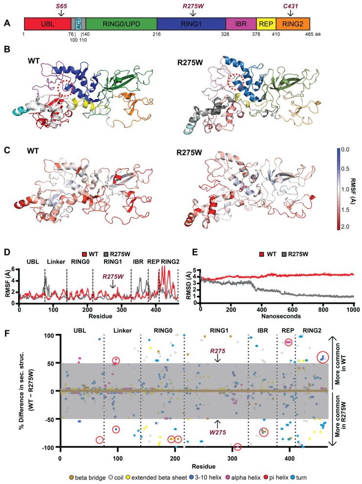
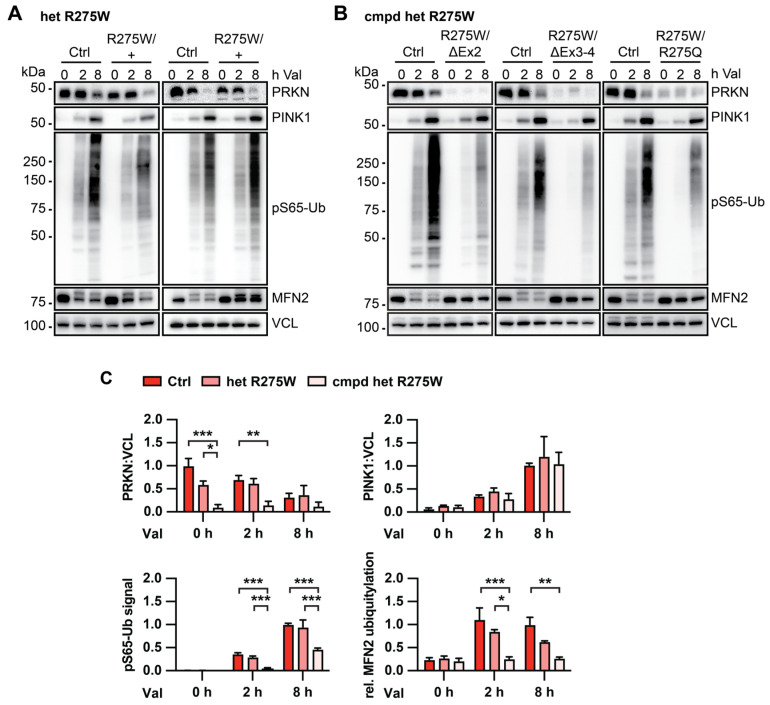
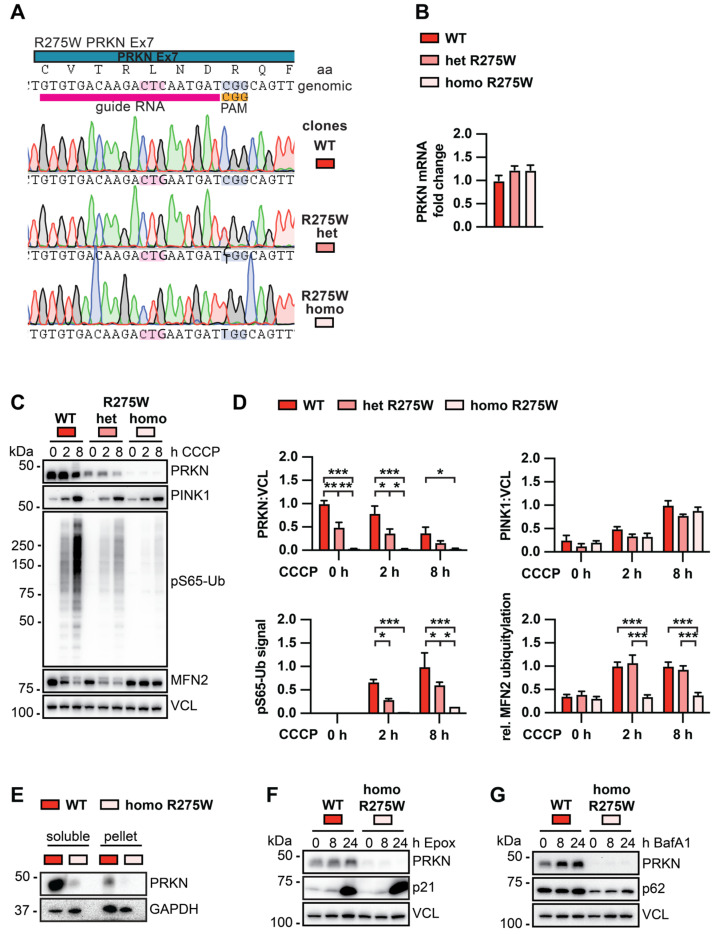
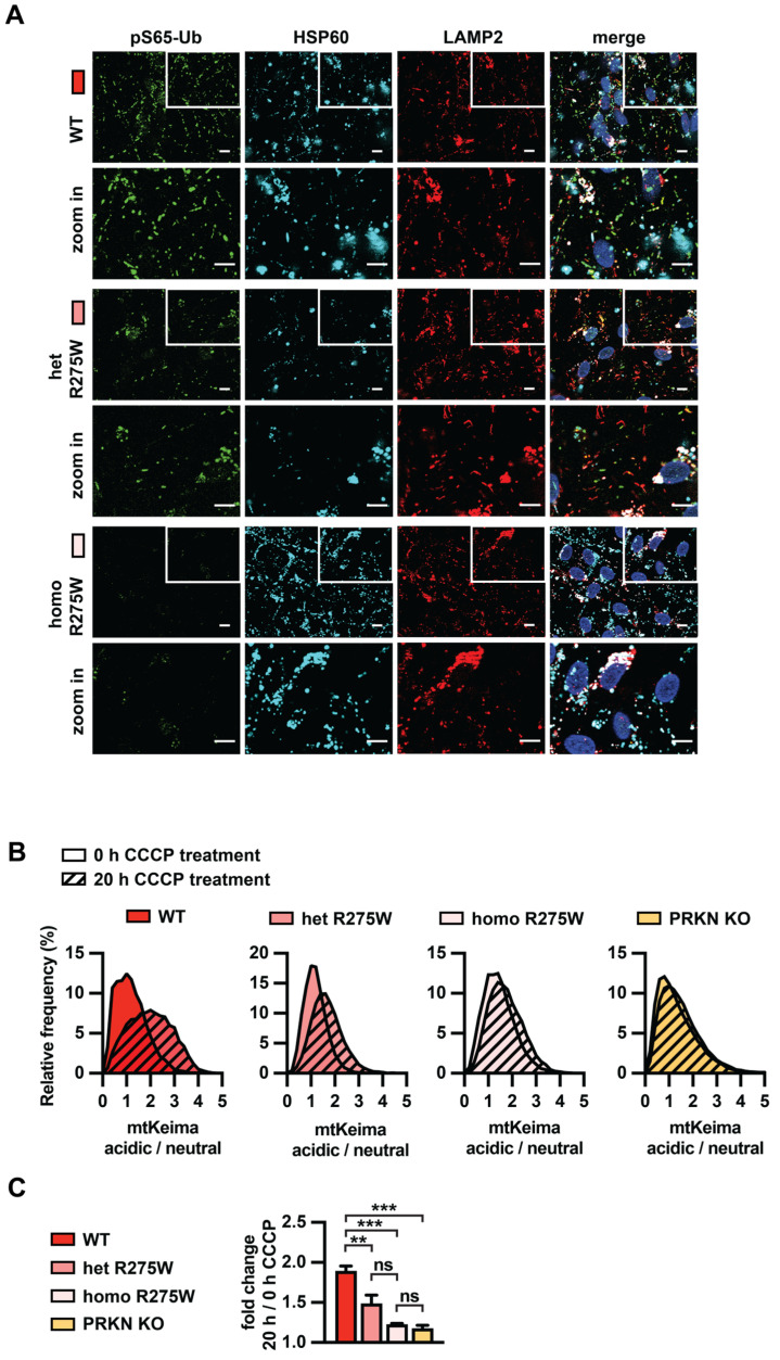
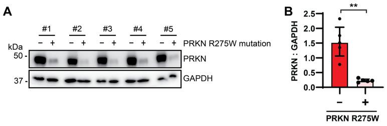
Mutations in the PINK1 and PRKN genes are the most frequent genetic cause of early-onset Parkinson disease. The pathogenic p.R275W substitution in PRKN is the most frequent substitution observed in patients, and thus far has been characterized mostly through overexpression models that suggest a possible gain of toxic misfunction. However, its effects under endogenous conditions are largely unknown. We used patient fibroblasts, isogenic neurons, and post-mortem human brain samples from carriers with and without PRKN p.R275W to assess functional impact. Immunoblot analysis and immunofluorescence were used to study mitophagy activation, and mitophagy execution was analyzed by flow cytometry of the reporter mitoKeima. The functional analysis was accompanied by structural investigation of PRKN p.R275W. We observed lower PRKN protein in fibroblasts with compound heterozygous p.R275W mutations. Isogenic neurons showed an allele-dose dependent decrease in PRKN protein. Lower PRKN protein levels were accompanied by diminished phosphorylated ubiquitin and decreased MFN2 modification. Mitochondrial degradation was also allele-dose dependently impaired. Consistently, PRKN protein levels were drastically reduced in human brain samples from p.R275W carriers. Finally, structural simulations showed significant changes in the closed form of PRKN p.R275W. Our data suggest that under endogenous conditions the p.R275W mutation results in a loss-of-function by destabilizing PRKN.

Keywords: PINK1; PRKN; Parkinson disease; mitophagy; parkin; ubiquitin.


Conflict of interest statement

Mayo Clinic, F.C.F. and W.S. hold a patent related to PRKN activators (Small Molecule Activators of Parkin Enzyme Function, US patent,11401255B2; 2 August 2022). D.P.N. receives research funding from Spark Therapeutics. All other authors declare they have no competing interests. Z.K.W. serves as PI or Co-PI on Biohaven Pharmaceuticals, Inc. (BHV4157-206), Vigil Neuroscience, Inc. (VGL101-01.002, VGL101-01.201, PET tracer development protocol, Csf1r biomarker and repository project, and ultra-high field MRI in the diagnosis and management of CSF1R-related adult-onset leukoencephalopathy with axonal spheroids and pigmented glia), and ONO-2808-03 projects/grants. He serves as Co-PI of the Mayo Clinic APDA Center for Advanced Research and as an external advisory board member for the Vigil Neuroscience, Inc., and as a consultant for Eli Lilly & Company and for NovoGlia, Inc. This research was conducted in compliance with Mayo Clinic conflict of interest policies.

Figures

Figure 1
Figure 1
Structure of PRKN WT and p.R275W and conformational changes. (A) Schematic overview of the PRKN protein structure and individual domains: Ub-like domain (UBL, red), linker domain (gray), activating element (ACT, cyan), really interesting new gene domain (RING0, green), RING1 (blue), in-between-RING domain (IBR, purple), repressor element of PRKN (REP, yellow), and RING2 (orange). (B) A 3D model of the autoinhibited structure of PRKN WT and p.R275W with individual domains colored as in (A); the location of position 275 is highlighted by a red dashed circle. (C) A 3D heat map illustration of the root mean square fluctuation (RMSF) for PRKN WT and p.R275W over the course of the simulations. (D) Comparison of RMSF for each residue of PRKN WT (red) and PRKN p.R275W (gray). Domains are labeled and separated with dotted lines. (E) Root mean square deviation (RMSD) of PRKN WT (red) and PRKN p.R275W (gray) structure over 1 µs simulation. (F) Per-residue difference plot of the secondary structure averaged over the entire simulation and colored by secondary structure feature. Shown are percent occupancy times of each residue in a specific secondary structure for PRKN WT (top) and PRKN p.R275W (bottom). Differences greater than 50% (outside the gray area) are considered most meaningful. Secondary structure colors are indicated at the bottom of the figure.
Figure 2
Figure 2
Biochemical analysis of PINK1-PRKN signaling in human fibroblasts with or without the PRKN p.R275W mutation. Dermal fibroblasts from controls and PD patients with heterozygous (A) or compound heterozygous (B) PRKN p.R275W mutation were treated with 1 μM valinomycin (Val) for 0, 2, or 8 h. Cell lysates were collected for Western blot analysis and probed with antibodies against PRKN, PINK1, pS65-Ub, and MFN2. VCL was used as loading control. (C) Densitometric analysis of PRKN and PINK1 Western blot signals normalized to VCL. Relative modification of MFN2 was calculated as the ratio of the upper (ubiquitylated) to lower (unmodified) MFN2 band. Quantification of pS65-Ub levels measured by sandwich ELISA. Data were normalized to control cells, either at the 0 or the 8 h time point. Data shown are mean ± SEM (n = 4 for WT, n = 2 for p.R275W heterozygous, and n = 3 for compound heterozygous). Statistical analysis was performed by two-way ANOVA followed by Tukey’s post hoc test (* p < 0.05, ** p < 0.01, *** p < 0.001). Only statistically significant comparisons are indicated.
Figure 3
Figure 3
Biochemical analysis of PINK1-PRKN signaling in isogenic DA neurons with or without the PRKN p.R275W mutation. (A) Schematic of CRISPR–Cas9 target location, gRNA, and results from Sanger sequencing of isogenic clones. (B) PRKN mRNA levels in WT, p.R275W heterozygous, and p.R275W homozygous gene-edited DA neurons. Data shown are mean ± SEM (n = 3). (C) WT, p.R275W heterozygous, and p.R275W homozygous gene-edited DA neurons were treated with 20 µM CCCP for 0, 2, or 8 h. Cell lysates were collected for Western blot analysis and probed with antibodies against PRKN, PINK1, pS65-Ub, MFN2, and VCL. Representative Western blot shows levels of PRKN, PINK1, pS65-Ub, and MFN2 for all three cell lines and treatments. (D) Densitometric analysis of PRKN and PINK1 normalized by VCL. Quantification of pS65-Ub protein levels by sandwich ELISA. Relative modification of MFN2 was calculated as the ratio of the upper (ubiquitylated) to lower (unmodified) MFN2 band. Statistical analysis was performed via two-way ANOVA (* p < 0.05, ** p < 0.01, *** p < 0.001) and data shown are mean ± SEM (n = 3). Only statistically significant comparisons are indicated. (E) Proteins from WT and PRKN p.R275W homozygous gene-edited DA neurons were sequentially extracted in two fractions for solubility analysis and probed with antibodies against PRKN and GAPDH. (F) Gene-edited DA WT neurons and PRKN p.R275W homozygous DA neurons were treated with 200 μM epoxomicin for 0, 8, or 24 h to test PRKN degradation via the proteasome. Cell lysates were collected for Western blot analysis and probed with antibodies against PRKN, p21, and VCL. (G) Gene-edited DA neurons were treated with 400 μM bafilomycin A1 for 0, 8, or 24 h to test for PRKN levels upon inhibition of the autophagosome-lysosome fusion. Cell lysates were collected and probed against PRKN, p62, and VCL.
Figure 4
Figure 4
Analysis of PINK1-PRKN mitophagy execution in p.R275W isogenic cell lines. (A) Immunofluorescence imaging for gene-edited DA neurons. WT, p.R275W hetero- or homozygous cells were treated with 20 µM CCCP for 8 h. Cells were stained with antibodies against pS65-Ub (green), lysosomal marker LAMP2 (red), and mitochondrial marker HSP60 (cyan). Hoechst was used to stain nuclei. Scale bar = 10 μm. (B) Quantification of mitophagy in WT, PRKN p.R275W heterozygous or homozygous neurons, and PRKN KO using flow cytometry of mitoKeima. Representative frequency distribution of acidic/neutral mitoKeima in untreated cells and the shift that is observed after 20 h CCCP treatment (shaded) are shown. (C) Fold change of the acidic/neutral mitoKeima ratio at 20 h CCCP treatment compared to untreated cells for each cell line. Data shown are mean ± SEM (n = 3). Statistical analysis was performed via one-way ANOVA (** p < 0.01, *** p < 0.001, ns = non-significant).
Figure 5
Figure 5
PRKN p.R275W levels are decreased in human post-mortem brain samples. (A) Age-, sex- and disease-matched human post-mortem brain samples were lysed for Western blot analysis and probed with antibodies against PRKN and GAPDH. (B) Densitometric analysis of PRKN normalized to GAPDH. Data shown are median ± interquartile range (IQR) (n = 5). Statistical analysis was performed using the Mann–Whitney test (** p < 0.01).

References

    1. Dickson D.W. Neuropathology of Parkinson disease. Park. Relat. Disord. 2018;46((Suppl. S1)):S30–S33. doi: 10.1016/j.parkreldis.2017.07.033. - DOI - PMC - PubMed
    1. Geisler S., Holmstrom K.M., Skujat D., Fiesel F.C., Rothfuss O.C., Kahle P.J., Springer W. PINK1/Parkin-mediated mitophagy is dependent on VDAC1 and p62/SQSTM1. Nat. Cell Biol. 2010;12:119–131. doi: 10.1038/ncb2012. - DOI - PubMed
    1. Narendra D.P., Jin S.M., Tanaka A., Suen D.F., Gautier C.A., Shen J., Cookson M.R., Youle R.J. PINK1 is selectively stabilized on impaired mitochondria to activate Parkin. PLoS Biol. 2010;8:e1000298. doi: 10.1371/journal.pbio.1000298. - DOI - PMC - PubMed
    1. Vives-Bauza C., Zhou C., Huang Y., Cui M., de Vries R.L., Kim J., May J., Tocilescu M.A., Liu W., Ko H.S., et al. PINK1-dependent recruitment of Parkin to mitochondria in mitophagy. Proc. Natl. Acad. Sci. USA. 2010;107:378–383. doi: 10.1073/pnas.0911187107. - DOI - PMC - PubMed
    1. Kane L.A., Lazarou M., Fogel A.I., Li Y., Yamano K., Sarraf S.A., Banerjee S., Youle R.J. PINK1 phosphorylates ubiquitin to activate Parkin E3 ubiquitin ligase activity. J. Cell Biol. 2014;205:143–153. doi: 10.1083/jcb.201402104. - DOI - PMC - PubMed

Publication types

MeSH terms

LinkOut - more resources