. 2024 Sep 20;13(18):1583.
doi: 10.3390/cells13181583.

A Flow Cytometry-Based Examination of the Mouse White Blood Cell Differential in the Context of Age and Sex

Affiliations

A Flow Cytometry-Based Examination of the Mouse White Blood Cell Differential in the Context of Age and Sex

Elise Arlt et al. Cells. .

Abstract

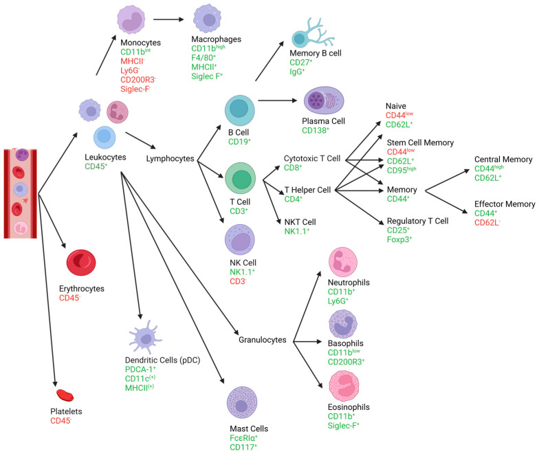
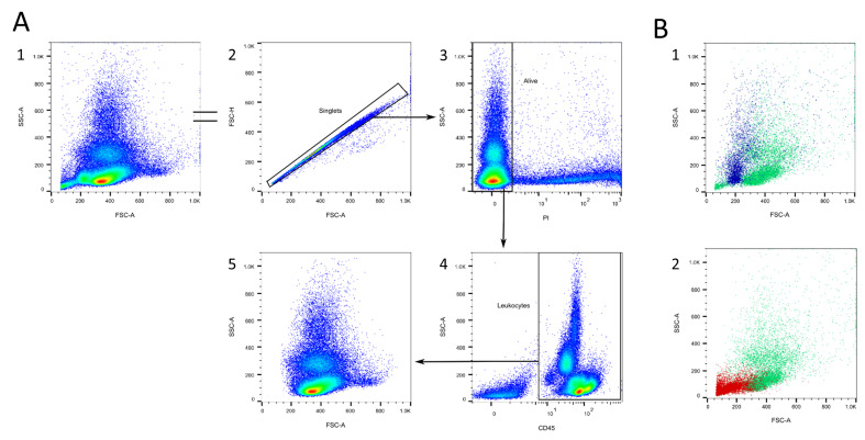
Analysis of the white blood cell differential as part of a flow cytometry-based approach is a common routine diagnostic tool used in clinics and research. For human blood, the methodological approach, suitable markers, and gating strategies are well-established. However, there is a lack of information regarding the mouse blood count. In this article, we deliver a fast and easy protocol for reprocessing mouse blood for the purpose of flow cytometric analysis, as well as suitable markers and gating strategies. We also present two possible applications: for the analysis of the whole blood count, with blood from a cardiac puncture, and for the analysis of a certain leukocyte subset at multiple time points in the framework of a mouse experiment, using blood from the facial vein. Additionally, we provide orientation values by applying the method to 3-month-old and 24-month-old male and female C57BL/6J mice. Our analyses demonstrate differences in the leukocyte fractions depending on age and sex. We discuss the influencing factors and limitations that can affect the results and that, therefore, need to be considered when applying this method. The present study fills the gap in the knowledge related to the rare information on flow cytometric analysis of mouse blood and, thus, lays the foundation for further investigations in this area.

Keywords: blood; blood sampling; immune cells; immune system; leukocyte subsets; leukocytes; mouse; mouse blood; mouse blood cells; reference values.


Conflict of interest statement

The authors declare no conflicts of interest. The funders had no role in the design of the study; in the collection, analyses, or interpretation of data; in the writing of the manuscript; or in the decision to publish the results.

Figures

Figure 1
Figure 1
The graphic shows the most important leukocyte subsets and their surface markers for identifying them. For each cell type, the corresponding pathway can be followed, based upon which the expressed markers are listed. This figure was designed on the basis of Section 3.1 “Gating strategies”. Created with BioRender.com.
Figure 2
Figure 2
First gating steps. (A) The doublets excluded (2). Dead cells are excluded with the help of propidium iodide (PI, 3). Then, the CD45+ cells are selected (4). (B) Dead cells accumulate as a small population in the FSC/SSC plot (blue, 1). Erythrocytes and platelets accumulate in the corner of the FSC/SSC plot (red, 2).
Figure 3
Figure 3
T cell gating. (A) CD3+ cells (T cells) are selected (2). T cells can further be classified by their expression in terms of CD4 and CD8 (3). CD4+CD25+FoxP3+ regulatory T cells can be defined (4). Both CD4+ and CD8+ T cells can further be classified by their expression in terms of CD95, CD44, and CD62L into naïve T cells, stem cell memory T cells (SCM), central memory T cells (CM), and effector memory T cells (EM, 5–8). (B) CD4+ T cells express higher CD3 levels compared to CD8+ T cells. (C) Sca-1 expression in CD4+ and CD8+ T cells. (D) Location of T cells (blue) in the FSC/SSC plot (1) and the CD45/SSC plot (2). Source of the plots: (A1–4) Panel 1; (A5–8,B,C) Panel 3.
Figure 4
Figure 4
B cell gating. (A) B cells can be marked with CD19 (2) and are MHCII+ IgM+ IgD+IgG (2,4,5). Most B cells express Sca-1 (6). Some of the B cells express CD138 or CD27 (3). (B) Location of B cells (violet) in the FSC/SSC plot (1) and the CD45/SSC plot (2). Source of the plots: (A) Preliminary experiments; (B) Panel 1.
Figure 5
Figure 5
NK cell gating. (A) NK cells are characterized by the expression of NK1.1 and the absence of CD3, while NKT cells express NK1.1, as well as CD3 (2). NK cells can be further subdivided into four maturation stages: CD11blow/−D27low/−, CD11blow/−CD27high/+, CD11bhigh/+CD27high/+, and CD11bhigh/+CD27low/− (3). The activity of NK cells can be analyzed by their expression in terms of activating NK cell receptors, as exemplarily shown for NCR1 and NKG2D (4+5). (B) Location of NK cells (orange) in the FSC/SSC (1) and the CD45/SSC plot (2). Source of the plots: Panel 2.
Figure 6
Figure 6
Monocyte and macrophage gating. (A) T, B, and NK cells are excluded in a lineage channel (2), then CD11b and CD11bhigh cells are excluded (3). Neutrophils are excluded via Ly6G (4), eosinophils via SSC (5), and basophils via CD200R3 (6). Monocytes can further be subdivided into a Ly6Chigh/+ and a Ly6Clow/− population (7). Ly6C MHCII+ monocyte-derived dendritic cells can be found (7). (B) Macrophages appear as a CD11bhigh Siglec-F+ F4/80+ MHCII+ Ly6G population (1–4). (C) Location of monocytes (pink) in the FSC/SSC plot (1) and the CD45/SSC plot (2). (D) Location of macrophages (pink) in the FSC/SSC plot (1) and the CD45/SSC plot (2). Source of the plots: (A,B3–4,C,D) Panel 4; (B1) Panel 6; (B2) preliminary experiments.
Figure 7
Figure 7
Granulocyte gating. (A) T, B, and NK cells are excluded in a lineage channel (2). Neutrophils can be determined as a CD11b+ Ly6Ghigh/+ Ly6Cint population (3 + 4). Eosinophils are characterized as CD11b+ Siglec-F+ Ly6C (3 + 5). Basophils are CD11bint CD200R3+Ly6C (6 + 7). (B) Location of neutrophils (blue), eosinophils (red), and basophils (pink) in the FSC/SSC plot (1,3,5) and the CD45/SSC plot (2,4,6). (C) After excluding CD11bhigh macrophages, granulocytes can roughly be subdivided in the CD45/SSC plot. Source of the plots: Panel 6.
Figure 8
Figure 8
Dendritic cell and mast cell gating. (A) T, B, and NK cells are excluded in a lineage channel (2). PDC are characterized as PDCA-1+ CD11cint MHCIIint (3 + 4). (B) T, B, and NK cells are excluded in a lineage channel (2). Mast cells can be determined as FcεRIα+CD117+ (3). (C) Location of DCs (pink) in the FSC/SSC plot (1) and the CD45/SSC plot (2). (D) Location of mast cells (red) in the FSC/SSC plot (1) and the CD45/SSC plot (2). Source of the plots: Panel 5.
Figure 9
Figure 9
Results from analyzing the blood from a cardiac puncture (Panel 1–6) to be used as orientation values (AY). The values for every leukocyte subset, as well as those subtypes that displayed differences between the groups, are presented. The data are presented as individual values and the mean. Groups: 3-month-old males (3 Mo M), 3-month-old females (3 Mo F), 24-months-old males (24 Mo M), and 24-month-old females (24 Mo F). Brackets indicate p values if lower than 0.1.
Figure 10
Figure 10
Gating strategy for Panel 7. B cells are identified with CD19 (2). T cells are CD3+ (2) and can be divided into CD4+, CD8+, and CD4CD8 populations (3). After excluding B and T cells, NK1.1+ NK cells can be distinguished (4). After excluding NK cells, monocytes can be roughly outlined with the help of CD11b and SSC (5). After excluding monocytes, different granulocyte populations can be roughly categorized by using SSC and CD45 (6).
Figure 11
Figure 11
Differences between cardiac (Panels 1–6) and facial vein blood sampling (Panel 7). The lines connect those values that belong to the same mouse.
Figure 12
Figure 12
Blood glucose values of all mice in the experiment. Two of the animals surpassed the set threshold of 11 mmol/L (dashed line) for stress hyperglycemia. Data are presented as individual values and the mean. Groups: 3-month-old males (3 Mo M), 3-month-old females (3 Mo F), 24-month-old males (24 Mo M), and 24-month-old females (24 Mo F). Brackets indicate p values.

Similar articles

Cited by

References

    1. Theml H., Diem H., Haferlach T. Taschenatlas der Hämatologie: Morphologische und klinische Diagnostik für die Praxis. Thieme; Stuttgart, NY, USA: 2002. 32 Tabellen, 5., vollst. überarb. Aufl.
    1. Christen U. Pathogen infection and autoimmune disease. Clin. Exp. Immunol. 2019;195:10–14. doi: 10.1111/cei.13239. - DOI - PMC - PubMed
    1. Rico L.G., Salvia R., Ward M.D., Bradford J.A., Petriz J. Flow-cytometry-based protocols for human blood/marrow immunophenotyping with minimal sample perturbation. STAR Protoc. 2021;2:100883. doi: 10.1016/j.xpro.2021.100883. - DOI - PMC - PubMed
    1. Dagur P.K., McCoy J.P. Collection, Storage, and Preparation of Human Blood Cells. Curr. Protoc. Cytom. 2015;73:5.1.1–5.1.16. doi: 10.1002/0471142956.cy0501s73. - DOI - PMC - PubMed
    1. Bundesinstitut für Risikobewertung Zahlen zu den 2021 in Deutschland Verwendeten Versuchstieren. 2022. [(accessed on 15 September 2024)]. Available online: https://www.bf3r.de/de/verwendung_von_versuchstieren_im_berichtsjahr_202....

Publication types

LinkOut - more resources