

Recent Progress in the Synthesis of 3D Complex Plasmonic Intragap Nanostructures and Their Applications in Surface-Enhanced Raman Scattering
- PMID: 39329807
- PMCID: PMC11430147
- DOI: 10.3390/bios14090433
Recent Progress in the Synthesis of 3D Complex Plasmonic Intragap Nanostructures and Their Applications in Surface-Enhanced Raman Scattering
Abstract
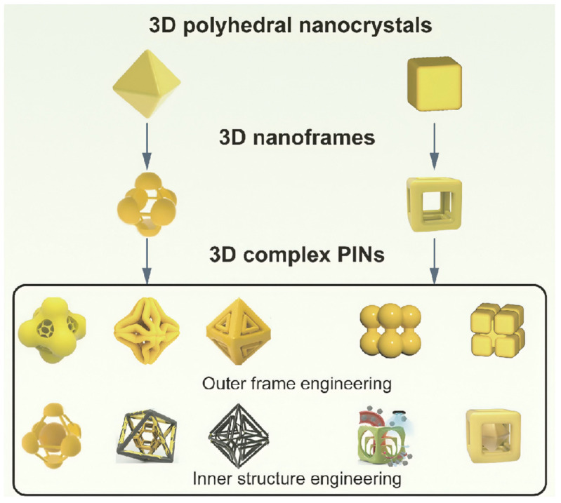
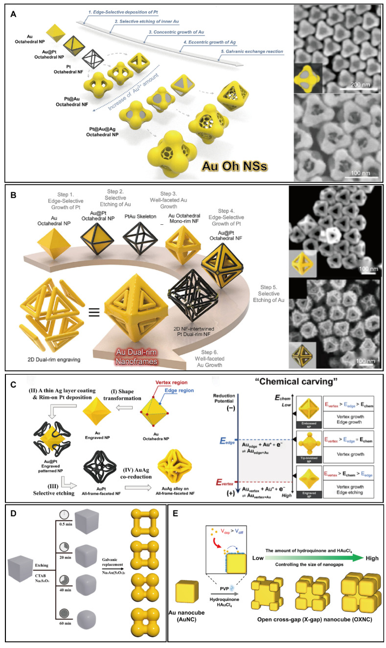
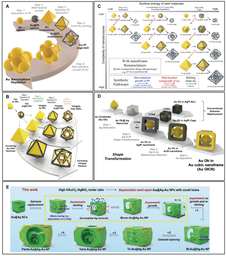
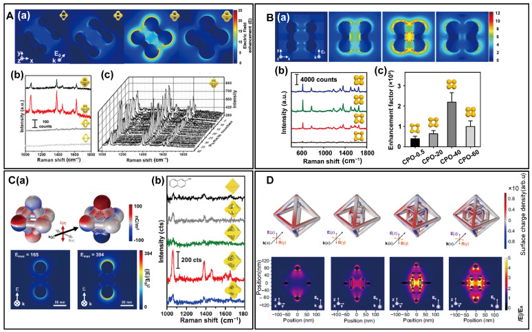
Plasmonic intragap nanostructures (PINs) have garnered intensive attention in Raman-related analysis due to their exceptional ability to enhance light-matter interactions. Although diverse synthetic strategies have been employed to create these nanostructures, the emphasis has largely been on PINs with simple configurations, which often fall short in achieving effective near-field focusing. Three-dimensional (3D) complex PINs, distinguished by their intricate networks of internal gaps and voids, are emerging as superior structures for effective light trapping. These structures facilitate the generation of hot spots and hot zones that are essential for enhanced near-field focusing. Nevertheless, the synthesis techniques for these complex structures and their specific impacts on near-field focusing are not well-documented. This review discusses the recent advancements in the synthesis of 3D complex PINs and their applications in surface-enhanced Raman scattering (SERS). We begin by describing the foundational methods for fabricating simple PINs, followed by a discussion on the rational design strategies aimed at developing 3D complex PINs with superior near-field focusing capabilities. We also evaluate the SERS performance of various 3D complex PINs, emphasizing their advanced sensing capabilities. Lastly, we explore the future perspective of 3D complex PINs in SERS applications.
Keywords: biosensing; intragap; localized surface plasmon resonance; nanostructure synthesis; near field; surface-enhanced Raman scattering.
Conflict of interest statement
The authors declare no conflicts of interest.
Figures


Similar articles
-
Hot spots in different metal nanostructures for plasmon-enhanced Raman spectroscopy.Nanoscale. 2013 Nov 21;5(22):10794-805. doi: 10.1039/c3nr02924g. Epub 2013 Oct 11. Nanoscale. 2013. PMID: 24113688
-
3D aluminum/silver hierarchical nanostructure with large areas of dense hot spots for surface-enhanced raman scattering.Electrophoresis. 2019 Dec;40(23-24):3123-3131. doi: 10.1002/elps.201900285. Epub 2019 Oct 14. Electrophoresis. 2019. PMID: 31576580
-
Nanoarchitecture Based SERS for Biomolecular Fingerprinting and Label-Free Disease Markers Diagnosis.Acc Chem Res. 2016 Dec 20;49(12):2725-2735. doi: 10.1021/acs.accounts.6b00384. Epub 2016 Dec 8. Acc Chem Res. 2016. PMID: 27993003 Free PMC article.
-
Toward Quantitative Surface-Enhanced Raman Scattering with Plasmonic Nanoparticles: Multiscale View on Heterogeneities in Particle Morphology, Surface Modification, Interface, and Analytical Protocols.J Am Chem Soc. 2022 Dec 14;144(49):22337-22351. doi: 10.1021/jacs.2c05950. Epub 2022 Dec 6. J Am Chem Soc. 2022. PMID: 36473154 Review.
-
Core-satellite nanostructures and their biomedical applications.Mikrochim Acta. 2022 Nov 24;189(12):470. doi: 10.1007/s00604-022-05559-0. Mikrochim Acta. 2022. PMID: 36435950 Review.
References
-
- Fleischmann M., Hendra P.J., McQuillan A.J. Raman Spectra of Pyridine Adsorbed at a Silver Electrode. Chem. Phys. Lett. 1974;26:163–166. doi: 10.1016/0009-2614(74)85388-1. - DOI
Publication types
MeSH terms
Grants and funding
- 22371127, 21902079, 62235008, 62288102/National Natural Science Foundation of China
- BK20212012/Natural Science Foundation of Jiangsu Province-Major Project
- GZR2022010028/Project of State Key Laboratory of Organic Electronics and Information Displays, Nanjing University of Posts and Telecommunications
LinkOut - more resources
Full Text Sources
Miscellaneous
