. 2024 Aug 24;46(9):9342-9358.
doi: 10.3390/cimb46090553.

The ER Stress Induced in Human Neuroblastoma Cells Can Be Reverted by Lumacaftor, a CFTR Corrector

Affiliations

The ER Stress Induced in Human Neuroblastoma Cells Can Be Reverted by Lumacaftor, a CFTR Corrector

Michela Pecoraro et al. Curr Issues Mol Biol. .

Abstract

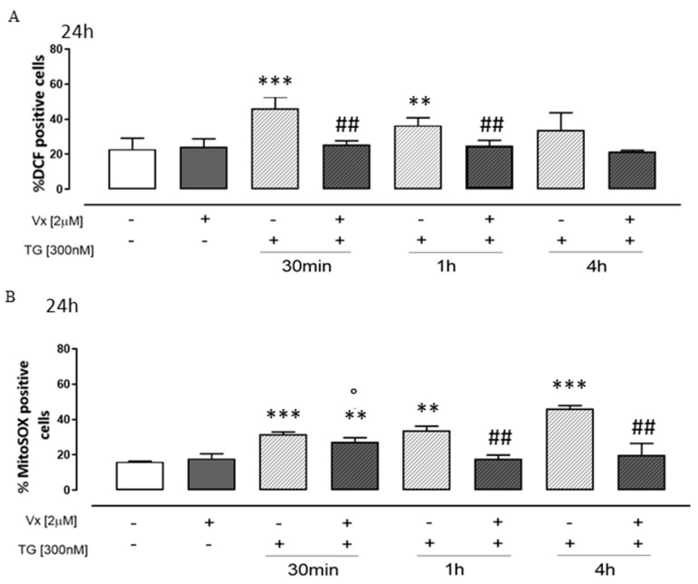
Most neurodegenerative diseases share a common etiopathogenesis, the accumulation of protein aggregates. An imbalance in homeostasis brought on by the buildup of misfolded proteins within the endoplasmic reticulum (ER) results in ER stress in the cell. Three distinct proteins found in the ER membrane-IRE1α, PERK, and ATF6-control the unfolded protein response (UPR), a signal transduction pathway that is triggered to restore normal physiological conditions. Buildup of misfolded proteins in ER lumen leads to a shunting of GRP78/BiP, thus triggering the UPR. PERK autophosphorylation leads to activation of ATF4, the transcription factor; finally, ATF6 activates the UPR's target genes, including GRP78/Bip. Accordingly, the UPR is a cellular reaction to an ER stress state that, if left unchecked for an extended period, results in apoptosis and irreversible damage. The identification of caspase 4, which is in the ER and is selectively activated by apoptotic stimuli caused by reticular stress, further demonstrated the connection between reticular stress and programed cell death. Moreover, oxidative stress and ER stress are linked. Oxidative stress is brought on by elevated quantities of radical oxygen species, both mitochondrial and cytosolic, that are not under the enzymatic regulation of superoxide dismutases, whose levels fall with increasing stress. Here, we evaluated the activity of Vx-809 (Lumacaftor), a drug used in cystic fibrosis, in SH-SY5Y neuronal cells, in which an ER stress condition was induced by Thapsigargin, to verify whether the drug could improve protein folding, suggesting its possible therapeutic use in proteinopathies, such as neurodegenerative diseases (NDs). Our data show that Vx-809 is involved in the significant reduction in protein produced under ER stress, particularly in the levels of Bip, ATF4, and ATF6 by Western blotting analysis, the reduction in ROS in the cytosol and mitochondria, and the reduction in the activation of the apoptotic pathway, measured by flow cytofluorimetry analysis and in restoring calcium homeostasis.

Keywords: ER stress; Lumacaftor; neuroblastoma cells; oxidative stress; protein misfolding.


Conflict of interest statement

The authors declare no conflicts of interest.

Figures

Figure 1
Figure 1
Schematic representation on the probable effect of Lumacaftor.
Figure 2
Figure 2
Vx-809 affects UPR activation. Cells were pretreated with 300 nM TG for 30 min or 2 or 4 h, to induce ER stress condition. Next, Vx-809 (2 µM) was added for 24 h. BiP (Panel (A)), ATF4 (Panel (C)), ATF6 (Panel (D)) and PERK/pPERK (Panel (E)) expressions in neuroblastoma cells were revealed by Western blotting analysis. Expression of actin or tubulin was employed as a loading control. Panel (B) illustrates the gel migration of the 452 bps unsplit fragment of XBP1 mRNA (XBP1 u) and the 426 bp split fragment (XBP1 s), achieved by RT-PCR. These results are presented as the average ± standard error of at least three separate, triplicate-performed studies. Data were processed according to the Mann–Whitney U-test. * p < 0.05, ** p < 0.005, and *** p < 0.001 vs. untreated cells; ° p < 0.05 and °° p < 0.005 vs. Vx-809-treated cells; # p < 0.05 vs. TG-treated cells.
Figure 2
Figure 2
Vx-809 affects UPR activation. Cells were pretreated with 300 nM TG for 30 min or 2 or 4 h, to induce ER stress condition. Next, Vx-809 (2 µM) was added for 24 h. BiP (Panel (A)), ATF4 (Panel (C)), ATF6 (Panel (D)) and PERK/pPERK (Panel (E)) expressions in neuroblastoma cells were revealed by Western blotting analysis. Expression of actin or tubulin was employed as a loading control. Panel (B) illustrates the gel migration of the 452 bps unsplit fragment of XBP1 mRNA (XBP1 u) and the 426 bp split fragment (XBP1 s), achieved by RT-PCR. These results are presented as the average ± standard error of at least three separate, triplicate-performed studies. Data were processed according to the Mann–Whitney U-test. * p < 0.05, ** p < 0.005, and *** p < 0.001 vs. untreated cells; ° p < 0.05 and °° p < 0.005 vs. Vx-809-treated cells; # p < 0.05 vs. TG-treated cells.
Figure 3
Figure 3
Vx-809 hampers calcium signaling. Cells were pretreated with 300 nM TG for 30 min or 1 or 4 h, to induce ER stress. Following this, Vx-809 (2 µM) was added for 24 h. After ER stress, the Vx-809 effect was determined on the reticular calcium pool in cells in calcium-free media in the presence of 1 nM TG (panel (A)), and intracellular calcium concentration was quantified using 1 μM of ionomycin (panel (B)). The results show the mean ± S.E.M. of the delta (δ) increase in the fluorescence of the FURA 2 ratio (340/380 nm) from a minimum of three separate experiments, each carried out in duplicate. The findings are presented as the average ± standard error of duplicate data from a minimum of three separate and identical tests. The Mann–Whitney U test analysis was performed on the data. ** p < 0.005 and *** p < 0.001 vs. untreated cells; ° p < 0.05 vs. Vx-809-treated cells; # p < 0.05 and ### p < 0.001 vs. TG-treated cells.
Figure 4
Figure 4
Vx-809 mitigates the oxidative damage caused by Thapsigargin. To cause ER stress, cells were pretreated with 300 nM TG for 30 min, 1 h, or 4 h. The corrector Vx-809 (2 µM) was then administered for a whole day. The probe 2′,7′-dichlorofluorescein diacetate (H2DCF-DA) was used to measure the amount of ROS produced in SH-SY5Y cells (Panel (A)). The percentage of DCF-positive cells in at least three independent experiments, each conducted in duplicate, was used to express the mean ± SEM of ROS generation. Using the MitoSOX Red probe, flow cytometry analysis was used to measure the amount of superoxide produced by the mitochondria in cells (Panel (B)). The expression of mitochondrial superoxide generation was calculated as the mean ± standard error of the proportion of cells positive for MitoSOX in three separate tests, each carried out in duplicate. SODIII expressions (Panel (C)) on neuroblastoma cells were detected by a Western blotting assay. Actin protein expression was used as loading control. The Mann–Whitney U test was used to evaluate the data. ** p < 0.005 and *** p < 0.001 vs. untreated cells; ° p < 0.05 vs. Vx-809-treated cells; # p < 0.05 and ## p < 0.005 vs. TG-treated cells.
Figure 4
Figure 4
Vx-809 mitigates the oxidative damage caused by Thapsigargin. To cause ER stress, cells were pretreated with 300 nM TG for 30 min, 1 h, or 4 h. The corrector Vx-809 (2 µM) was then administered for a whole day. The probe 2′,7′-dichlorofluorescein diacetate (H2DCF-DA) was used to measure the amount of ROS produced in SH-SY5Y cells (Panel (A)). The percentage of DCF-positive cells in at least three independent experiments, each conducted in duplicate, was used to express the mean ± SEM of ROS generation. Using the MitoSOX Red probe, flow cytometry analysis was used to measure the amount of superoxide produced by the mitochondria in cells (Panel (B)). The expression of mitochondrial superoxide generation was calculated as the mean ± standard error of the proportion of cells positive for MitoSOX in three separate tests, each carried out in duplicate. SODIII expressions (Panel (C)) on neuroblastoma cells were detected by a Western blotting assay. Actin protein expression was used as loading control. The Mann–Whitney U test was used to evaluate the data. ** p < 0.005 and *** p < 0.001 vs. untreated cells; ° p < 0.05 vs. Vx-809-treated cells; # p < 0.05 and ## p < 0.005 vs. TG-treated cells.
Figure 5
Figure 5
Vx-809 alters the process of apoptosis. Neuroblastoma cells were pretreated with 300 nM TG for 30 min, 2 h, or 4 h, to induce ER stress. Subsequently, 2 µM of the Vx-809 was administered for 24 h. After, the cells were stained by propidium iodide, and the fluorescence of individual nuclei was evaluated by flow cytometry (Panel (A)). The data are presented as the mean ± standard error of the percentage of hypodiploid nuclei from a minimum of three independent tests, each carried out in duplicate. The percentage of caspase 4 positive cells (Panel (B)) from at least three separate experiments, each carried out in duplicate, was given as mean ± S.E.M. The Mann–Whitney U test was utilized for data analysis. * p < 0.05, ** p < 0.05 and *** p < 0.001 vs. non-treated cells; ° p < 0.05 vs. Vx-809-treated cells; ## p < 0.005 and ### p < 0.001 vs. TG-treated cells.

Similar articles

Cited by

References

    1. Oakes S.A., Papa F.R. The role of endoplasmic reticulum stress in human pathology. Annu. Rev. Pathol. 2015;10:173–194. doi: 10.1146/annurev-pathol-012513-104649. - DOI - PMC - PubMed
    1. Rebecca B., Berlow H., Dyson J., Peter E. Wright, Expanding the Paradigm: Intrinsically Disordered Proteins and Allosteric Regulation. J. Mol. Biol. 2018;430:2309–2320. - PMC - PubMed
    1. Soto C., Pritzkow S. Protein misfolding, aggregation, and conformational strains in neurodegenerative diseases. Nat. Neurosci. 2018;21:1332–1340. doi: 10.1038/s41593-018-0235-9. - DOI - PMC - PubMed
    1. Ghemrawi R., Khair M. Endoplasmic Reticulum Stress and Unfolded Protein Response in Neurodegenerative Diseases. Int. J. Mol. Sci. 2020;21:6127. doi: 10.3390/ijms21176127. - DOI - PMC - PubMed
    1. Ashraf G., Greig N., Khan T., Hassan I., Tabrez S., Shakil S., Sheikh I., Zaidi S., Akram M., Jabir N., et al. Protein Misfolding and Aggregation in Alzheimer’s Disease and Type 2 Diabetes Mellitus. CNS Neurol. Disord. Drug Targets. 2014;13:1280–1293. doi: 10.2174/1871527313666140917095514. - DOI - PMC - PubMed

LinkOut - more resources