. 2024 Sep 12;46(9):10087-10111.
doi: 10.3390/cimb46090602.

Impacts of DROSHA (rs10719) and DICER (rs3742330) Variants on Breast Cancer Risk and Their Distribution in Blood and Tissue Samples of Egyptian Patients

Affiliations

Impacts of DROSHA (rs10719) and DICER (rs3742330) Variants on Breast Cancer Risk and Their Distribution in Blood and Tissue Samples of Egyptian Patients

Aly A M Shaalan et al. Curr Issues Mol Biol. .

Abstract

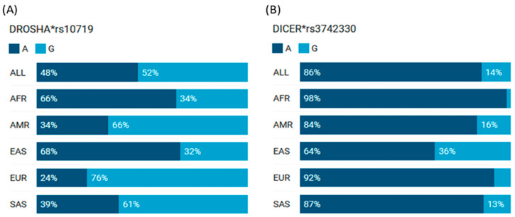
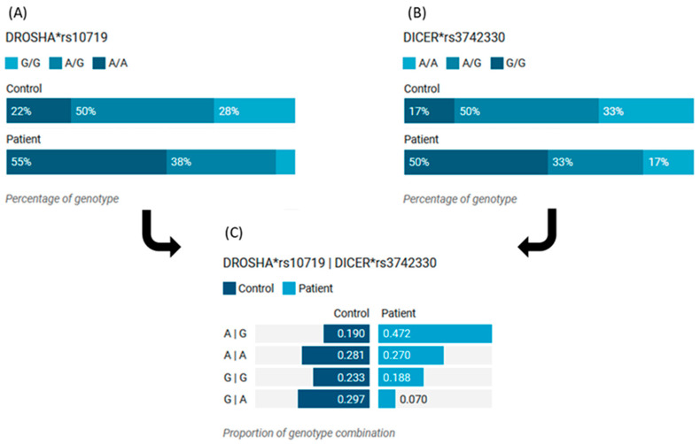
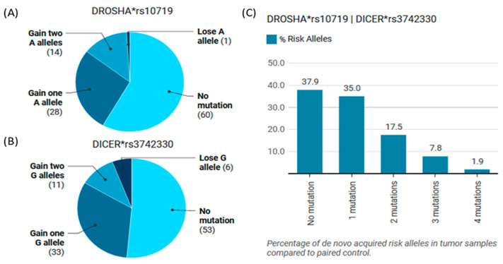
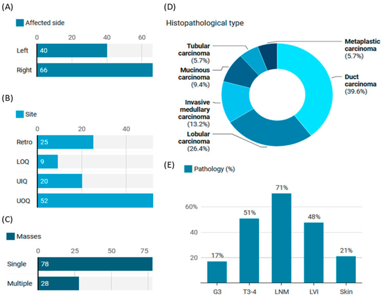
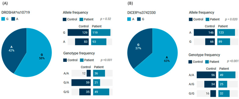
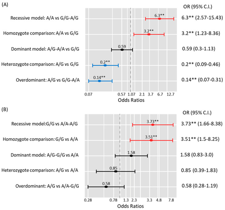
MicroRNAs (miRNAs) are small, noncoding RNAs that regulate gene expression and play critical roles in tumorigenesis. Genetic variants in miRNA processing genes, DROSHA and DICER, have been implicated in cancer susceptibility and progression in various populations. However, their role in Egyptian patients with breast cancer (BC) remains unexplored. This study aims to investigate the association of DROSHA rs10719 and DICER rs3742330 polymorphisms with BC risk and clinical outcomes. This case-control study included 209 BC patients and 106 healthy controls. Genotyping was performed using TaqMan assays in blood, tumor tissue, and adjacent non-cancerous tissue samples. Associations were analyzed using logistic regression and Fisher's exact test. The DROSHA rs10719 AA genotype was associated with a 3.2-fold increased risk (95%CI = 1.23-9.36, p < 0.001), and the DICER rs3742330 GG genotype was associated with a 3.51-fold increased risk (95%CI = 1.5-8.25, p = 0.001) of BC. Minor allele frequencies were 0.42 for rs10719 A and 0.37 for rs3742330 G alleles. The risk alleles were significantly more prevalent in tumor tissue than adjacent normal tissue (rs10719 A: 40.8% vs. 0%; rs3742330 G: 42.7% vs. 0%; p < 0.001). However, no significant associations were observed with clinicopathological features or survival outcomes over a median follow-up of 17 months. In conclusion, DROSHA rs10719 and DICER rs3742330 polymorphisms are associated with increased BC risk and more prevalent in tumor tissue among our cohort, suggesting a potential role in miRNA dysregulation during breast tumorigenesis. These findings highlight the importance of miRNA processing gene variants in BC susceptibility and warrant further validation in larger cohorts and different ethnic populations.

Keywords: DICER; DROSHA; breast cancer; microRNA; polymorphism; susceptibility.


Conflict of interest statement

The authors declare no conflicts of interest.

Figures

Figure 1
Figure 1
The allele frequencies of DROSHA rs10719 and DICER rs3742330 variants in different populations. (A) DROSHA (B) DICER. AMR: American; AFR: African; EAS: East Asian; EUR: European; SAS: South Asians. Data source: 1000 Genomes Project Phase 3 allele frequencies [Ensembl.org] (last accessed on 20 March 2024)”. *rs: reference sequence.
Figure 2
Figure 2
Analysis of the DROSHA gene structure and its associated 3D interactions with other genes and variants mediated by chromatin loops. (A) The DROSHA gene is on the short arm of chromosome 5 on the forward strand, following the ‘GRCh38.p14’ assembly. The rs10719 variant is positioned at 5:31,401,340 (highlighted), where the ancestral nucleotide ‘A’ is replaced by the alternative (minor) allele ‘G’ (https://www.ncbi.nlm.nih.gov/snp/rs10719). (B) A Circos plot illustrating the chromatin loops and other 2D characteristics related to the variant of interest, generated using 3DSNP 2.0 (https://omic.tech/3dsnpv2/). The plot displays, from the outer edge to the inner section, the chromatin states, annotated genes, the current SNP of interest and associated SNPs, and 3D chromatin interactions. A color key corresponding to the chromatin states and loops for twelve distinct cell types has been detailed previously [23]. (C) DROSHA’s subcellular localization can be accessed via https://www.proteinatlas.org/ENSG00000113360-DROSHA/subcellular. (D) The conservation score for the variant of interest is recorded as 2.277, derived from multiple alignments of vertebrate (n = 46) and mammalian (n = 33) genomes. All databases were last accessed on 30 March 2024.
Figure 3
Figure 3
Structural analysis of the DICER gene and its associated 3D interactions with other genes and variants mediated by chromatin loops. (A) The DICER gene is situated on the long arm of chromosome 14 on the reverse strand, aligning with the ‘GRCh38.p14’ assembly. The rs3742330 variant is found at position 14: 95,087,025 (highlighted), where the ancestral nucleotide ‘A’ is replaced by the alternative (minor) allele ‘G’ (https://www.ncbi.nlm.nih.gov/snp/rs3742330). (B) A Circos plot displaying the chromatin loops and other 2D features related to the variant of interest was created using “3DSNP 2.0 (https://omic.tech/3dsnpv2/)”. The plot illustrates, from the outermost section to the inner, the chromatin states, annotated genes, the currently examined SNP and its associated SNPs, and 3D chromatin interactions. The color key for chromatin states and loops across twelve different cell types is provided in a previous work [23]. (C) Information regarding the subcellular distribution of DICER can be accessed at https://www.proteinatlas.org/ENSG00000100697-DICER1/subcellular. (D) The conservation score for the variant of interest is reported as −0.023, derived from multiple alignments of vertebrate (n = 46) and mammalian (n = 33) genomes. All databases were last accessed on 30 March 2024.
Figure 4
Figure 4
Genotype combination analysis of DICER and DROSHA genes in tissues of BC women. (A) Distribution of DROSHA genotypes in patients and controls. (B) Distribution of DICER genotypes in patients and controls. (C) Distribution of combined DROSHA and DICER genotypes in patients and controls. *rs: reference sequence.
Figure 5
Figure 5
Somatic mutation analyses of DICER and DROSHA polymorphisms in paired tissues of women with BC. (A) Genotype alteration of the DROSHA gene in cancer and non-cancer tissues. The A allele is considered the risky variant. (B) Genotype alteration of DICER gene in cancer and non-cancer tissues. G allele is considered the risky variant. (C) Genotype alteration of combined DROSHA and DICER genes in cancer/non-cancer tissues. *rs: reference sequence.
Figure 6
Figure 6
Clinical presentation of BC women with blood samples (n = 106). (A) Counts of affected sides. (B) Counts of affected locations. (C) Counts of patients according to the number of masses at the time of presentation. (D) The different histopathological types. (E)Pathology-related data of patients with BC and provided blood samples. G3: Grade 3, T3-4: stage 3-4, LNM: Lymph node metastasis, LVI: lymphovascular infiltration.
Figure 7
Figure 7
The genotype and allele frequencies of the DICER and DROSHA genes in blood samples of BC and non-cancer women: (A) DROSHA rs10719; (B) DICER rs3742330. Pie charts represented the percentage of each allele in the overall cohorts (cases and controls). The bar chart showed the frequencies (counts) of cohorts per allele or genotype. A two-sided Chi-square test was used. Significance was set at p < 0.05. *rs: reference sequence.
Figure 8
Figure 8
Genetic association models for disease risk assessment: (A) DROSHA rs10719; (B) DICER rs3742330. Multivariate regression models were performed and shown as odds ratios (ORs) and 95% confidence intervals (95%CI). Adjusted variables were patient age at diagnosis, marital status, occupation, a family history of cancer, prior breast problems, smoking, body mass index, diabetes, hypertension, and hepatitis C virus infection. The red line is a risky genotype, the blue line is a protective genotype, and the black line is insignificant. ** indicate significance at p-value < 0.05.

Similar articles

References

    1. Wilkinson L., Gathani T. Understanding breast cancer as a global health concern. Br. J. Radiol. 2022;95:20211033. doi: 10.1259/bjr.20211033. - DOI - PMC - PubMed
    1. Bray F., Laversanne M., Sung H., Ferlay J., Siegel R.L., Soerjomataram I., Jemal A. Global cancer statistics 2022: GLOBOCAN estimates of incidence and mortality worldwide for 36 cancers in 185 countries. CA A Cancer J. Clin. 2024;74:229–263. doi: 10.3322/caac.21834. - DOI - PubMed
    1. Buocikova V., Rios-Mondragon I., Pilalis E., Chatziioannou A., Miklikova S., Mego M., Pajuste K., Rucins M., Yamani N.E., Longhin E.M., et al. Epigenetics in Breast Cancer Therapy-New Strategies and Future Nanomedicine Perspectives. Cancers. 2020;12:3622. doi: 10.3390/cancers12123622. - DOI - PMC - PubMed
    1. Sarvari P., Ramírez-Díaz I., Mahjoubi F., Rubio K. Advances of Epigenetic Biomarkers and Epigenome Editing for Early Diagnosis in Breast Cancer. Int. J. Mol. Sci. 2022;23:9521. doi: 10.3390/ijms23179521. - DOI - PMC - PubMed
    1. Plawgo K., Raczynska K.D. Context-Dependent Regulation of Gene Expression by Non-Canonical Small RNAs. Noncoding RNA. 2022;8:29. doi: 10.3390/ncrna8030029. - DOI - PMC - PubMed

LinkOut - more resources