Review
. 2024 Aug 23;10(9):547.
doi: 10.3390/gels10090547.

Natural Regenerative Hydrogels for Wound Healing

Affiliations
Review

Natural Regenerative Hydrogels for Wound Healing

Mariana Chelu et al. Gels. .

Abstract

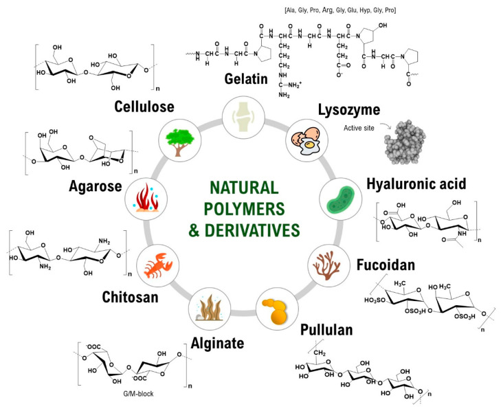
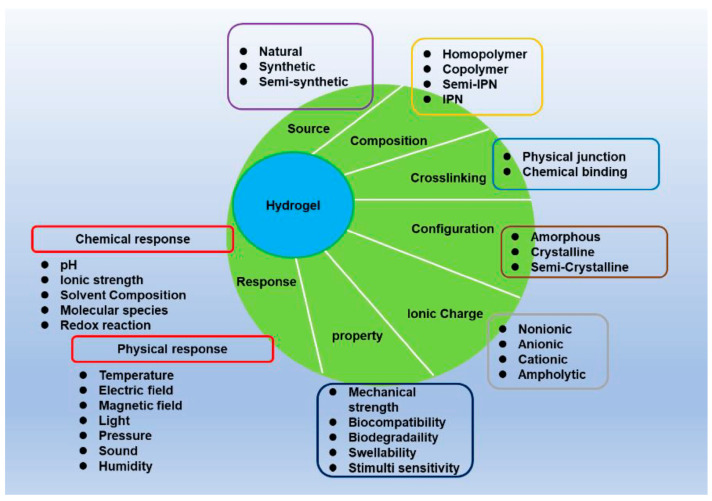
Regenerative hydrogels from natural polymers have come forth as auspicious materials for use in regenerative medicine, with interest attributed to their intrinsic biodegradability, biocompatibility, and ability to reassemble the extracellular matrix. This review covers the latest advances in regenerative hydrogels used for wound healing, focusing on their chemical composition, cross-linking mechanisms, and functional properties. Key carbohydrate polymers, including alginate, chitosan, hyaluronic acid, and polysaccharide gums, including agarose, carrageenan, and xanthan gum, are discussed in terms of their sources, chemical structures and specific properties suitable for regenerative applications. The review further explores the categorization of hydrogels based on ionic charge, response to physiological stimuli (i.e., pH, temperature) and particularized roles in wound tissue self-healing. Various methods of cross-linking used to enhance the mechanical and biological performance of these hydrogels are also examined. By highlighting recent innovations and ongoing challenges, this article intends to give a detailed understanding of natural hydrogels and their potential to revolutionize regenerative medicine and improve patient healing outcomes.

Keywords: antibacterial activity; biocompatibility; biodegradability; cross-linking mechanisms; extracellular matrix; glycosaminoglycans; polysaccharide gums; regenerative hydrogels; tissue engineering; wound healing.


Conflict of interest statement

The authors declare no conflicts of interest.

Figures

Figure 1
Figure 1
Natural carbohydrate polymers used for natural hydrogels [4].
Figure 2
Figure 2
The main characteristics of hydrogels [14].
Figure 3
Figure 3
Schemes showing (A) the preparation of APZC dressings and (B) the antibacterial activity [52].
Figure 4
Figure 4
(I) Aloe vera-based dressing: (a) dry and (b) wet hydrogel structure. Skin wound with time (c) 0 min; (d) 5 min; (e) 20 days; (f,g) images showing inverted vials with AV hydrogels. (II) Fluorescent images for dead and live L929 cells (A) control; (BF) AV5 treated; (GK) AV10 treated (48 h). Hydrogel concentrations are (B,G) 10 mg/mL; (C,H) 25 mg/mL; (D,I) 50 mg/mL; (E,J) 75 mg/mL; (F,K)100 mg/mL. (III) Healing after wound generation in vitro (24 h): (a) optical micrographs. (b) closure percentage [106].
Figure 5
Figure 5
(I) PEG-GAG hydrogels implanted subdermally. (A) Scheme of the implantation site: Dermis (De), Adipose Tissue (AdTi), Skeletal muscle (SkMu), Hypodermis (HyDe), Capsule (Cap), Implant (Imp). (B) Foreign body reaction thickness, ns—not significant, ***—p ≤ 0.001. (C) Immunostaining after 28 days. Scale bar 200 μm. (II) Angiogenic effects, hydrogels loaded with different cytokines. (A) CD31 immunostaining of hydrogels (dark blue) with vascular structures (brown). Scale bar 200 μm. (B) Vessel formation [125], ns—not significant, ***—p ≤ 0.001.
Figure 6
Figure 6
(I) Optical image of the reinforced hydrogel. (II) (a) Image of the heart before explantation showing the white patch, the pink ischemic region and the coronary artery ligature. (b) Image of the heart after explantation, showing the ischemic region. Images of the transversal section through the ventricle wall, post-implantation, for (c) the control case without growth factors, and (d) case treated with HA:Hp-silk patch containing growth factors, showing a reduced damaged region, nearly full wall thickness, and significantly less fibrous deposition [126].
Figure 7
Figure 7
(A) Computed tomography 3D images of knee joints treated 50 days with hydrogels, area (B) and volume (C) of osteophytes. * p < 0.05, ** p < 0.01, *** p < 0.001, n = 5. (DH) Safranin staining of the paraffin sections of knee joints. An enlarged view is shown at the bottom of each image. (I) OARSI score of sections stained with safranin O-fast green. * p < 0.05, *** p < 0.001 and **** p < 0.0001, n = 5 [127].
Figure 8
Figure 8
Hydrogel scaffold: (A) before; (B) after lyophilization. Three-dimensional printed scaffold: (C) before; (D) after lyophilization [135].
Figure 9
Figure 9
Image of hydrogels combining silk fibroin and tyramine-substituted gelatin with the human placental extracellular matrix, showing changes in transparency according to composition [138].
Figure 10
Figure 10
Three-dimensional printed SGE hydrogel containing human fibroblasts. (A): (i) scheme of the 3D printing process showing the combination of cross-linking agents in the support bath. (ii) Scaffold printed into the support bath and (iii) after washing. (B): Different shapes 3D printed with SGE hydrogel. (CE) (i): printed scaffolds. (CE) (ii): staining of live and dead cells after one and seven days. (E) (iii): Metabolic activity increases over time (p < 0.05, Alamar Blue staining, *** indicates significant differences of p  <  0.05 between Day 7 and Day 1) [138].
Figure 11
Figure 11
(I) Optical micrographs of GRH gelatin-based hydrogel scaffolds: (A) wet; (B) lyophilized and SEM micrographs (C) 50×; (D) 1000×. (II) (A) Scheme of wound evolution evaluation. (B) Optical images of wounds treated with GRH for 13 days. Scale bar: 2 cm. (C) Reduction of the wound sizes (% of the initial wound size, n = 6) [139].

References

    1. Gomez-Florit M., Pardo A., Domingues R.M.A., Graça A.L., Babo P.S., Reis R.L., Gomes M.E. Natural-Based Hydrogels for Tissue Engineering Applications. Molecules. 2020;25:5858. doi: 10.3390/molecules25245858. - DOI - PMC - PubMed
    1. Revete A., Aparicio A., Cisterna B.A., Revete J., Luis L., Ibarra E., Segura González E.A., Molino J., Reginensi D. Advancements in the Use of Hydrogels for Regenerative Medicine: Properties and Biomedical Applications. Int. J. Biomater. 2022;1:3606765. doi: 10.1155/2022/3606765. - DOI - PMC - PubMed
    1. Yang Y., Xu L., Wang J., Meng Q., Zhong S., Gao Y., Cui X. Recent advances in polysaccharide-based self-healing hydrogels for biomedical applications. Carbohydr. Polym. 2022;283:119161. doi: 10.1016/j.carbpol.2022.119161. - DOI - PubMed
    1. Silva A.C.Q., Silvestre A.J.D., Vilela C., Freire C.S.R. Natural Polymers-Based Materials: A Contribution to a Greener Future. Molecules. 2022;27:94. doi: 10.3390/molecules27010094. - DOI - PMC - PubMed
    1. Luo M., Zhang X., Wu J., Zhao J. Modifications of polysaccharide-based biomaterials under structure-property relationship for biomedical applications. Carbohydr. Polym. 2021;266:118097. doi: 10.1016/j.carbpol.2021.118097. - DOI - PubMed

LinkOut - more resources