. 2024 Sep 2;10(9):571.
doi: 10.3390/gels10090571.

Papain Injection Creates a Nucleotomy-like Cavity for Testing Gels in Intervertebral Discs

Affiliations

Papain Injection Creates a Nucleotomy-like Cavity for Testing Gels in Intervertebral Discs

Jan Ulrich Jansen et al. Gels. .

Abstract

Biomaterials, such as hydrogels, have an increasingly important role in the development of regenerative approaches for the intervertebral disc. Since animal models usually resist biomaterial injection due to high intradiscal pressure, preclinical testing of the biomechanical performance of biomaterials after implantation remains difficult. Papain reduces the intradiscal pressure, creates cavities within the disc, and allows for biomaterial injections. But papain digestion needs time, and cadaver experiments that are limited to 24 h for measuring range of motion (ROM) cannot not be combined with papain digestion just yet. In this study, we successfully demonstrate a new organ culture approach, facilitating papain digestion to create cavities in the disc and the testing of ROM, neutral zone (NZ), and disc height. Papain treatment increased the ROM by up to 109.5%, extended NZ by up to 210.9%, and decreased disc height by 1.96 ± 0.74 mm. A median volume of 0.73 mL hydrogel could be injected after papain treatment, and histology revealed a strong loss of proteoglycans in the remaining nucleus tissue. Papain has the same biomechanical effects as known from nucleotomies or herniations and thus creates a disc model to study such pathologies in vitro. This new model can now be used to test the performance of biomaterials.

Keywords: biomechanics; chemonucleolysis; disc height; nucleus replacement; organ culture; papain; range of motion.


Conflict of interest statement

The authors declare no conflicts of interest.

Figures

Figure 1
Figure 1
Papain injection and its macroscopic effects. (a,b) Embedded bovine tail functional spinal units in cell culture medium in sterile container. (a) shows papain with contrast agent after injection in the intervertebral disc (IVD) center and (b) shows state after 24 h incubation at 37 °C. (c) Transversal X-ray of cavity 7 days after the papain injection. (dg) Transversal views of dissected discs on day 7 for papain group. (hk) Transversal views of dissected discs on day 7 for sham group.
Figure 2
Figure 2
Flexibility. Mean range of motion (ROM) and mean neutral zone (NZ) with standard deviation in three motion planes for the intact condition (n = 36) and subsequent time points after−incubation and after−loading for specimens either treated with papain (n = 21) or phosphate-buffered saline (PBS) as sham (n = 15). Significant differences between the intact and the after−incubation state: $ p < 0.05, $$ p < 0.01, $$$ p < 0.001. Significant differences between the after−incubation and the after−loading state: ### p < 0.001. Significant differences between the groups for each time point; ** p < 0.01, *** p < 0.001.
Figure 3
Figure 3
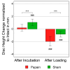
Disc height change. Plots of mean disc height change with standard deviation in mm for the time points after−incubation and after−loading normalized to the intact condition for specimens either treated with papain (n = 21) or PBS as sham (n = 15). Significant differences between the intact and the after−incubation states: $$$ p < 0.001. Significant differences between the after−incubation and the after−loading states: ### p < 0.001. Significant differences between the groups for each time point: *** p < 0.001.
Figure 4
Figure 4
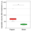
Injectable volume after treatment. Boxplots of injectable volume of hydrogel after incubation and loading in µL for specimens either treated with papain (n = 6) or PBS as sham (n = 6). Asterisks indicate significant differences between the groups. Level of significance: ** p < 0.01. Outliers: °.
Figure 5
Figure 5
Effects of papain digestion and complex loading on matrix composition and tissue integrity. (a) Histological overview of representative half IVDs of the bovine tail (n = 6) for papain and sham. Safranin-O/Fast-Green colors proteoglycan-rich tissue red and other tissue, such as collagen, green. (b) Sulphated glycosaminoglycan content quantified at the end of the experiment on day 7 for papain and sham group. n = 6 tissue punches were taken from the nucleus pulposus (NP) and anulus fibrosus (AF). The sulphated glycosaminoglycan content was normalized to the dry weight of the punched tissue. (c) Histological staining of sham discs (left column) without complex loading versus sham discs (right column) with complex loading on day 7. Sagittal IVD sections were stained with Safranin-O/Fast-Green for visualization of the tissue. The upper row shows tissue from the AF whereas the lower row shows tissue from the transition zone (TZ) (scale bar, 500 µm). Arrows indicate exemplary micro-damage within the tissue.
Figure 6
Figure 6
Method and machines. (a) Fresh bovine tail segment intact with antero-posterior X-ray. (b) Preparation and embedding of mono-segmental specimen with flanges and motion tracking markers. (c) Universal spine tester (Wilke et al., 1994 [88]) used for testing ROM and NZ with pure moments in three motion planes. (d) Universal testing machine for compression test to measure disc height change. (e) Injection of either papain or PBS as sham under sterile conditions. (f) Incubation in cell culture medium in hypoxic environment at 37 °C. (g) Dynamic disc loading simulator (Wilke et al., 2016 [89]) for applying complex loading for 2700 cycles. (h) Double chamber syringe for injecting radiopaque hydrogel for determining the injectable volume.

References

    1. Vos T., Flaxman A.D., Naghavi M., Lozano R., Michaud C., Ezzati M., Shibuya K., Salomon J.A., Abdalla S., Aboyans V., et al. Years lived with disability (YLDs) for 1160 sequelae of 289 diseases and injuries 1990-2010: A systematic analysis for the Global Burden of Disease Study 2010. Lancet. 2012;380:2163–2196. doi: 10.1016/S0140-6736(12)61729-2. - DOI - PMC - PubMed
    1. Hoy D., March L., Brooks P., Blyth F., Woolf A., Bain C., Williams G., Smith E., Vos T., Barendregt J., et al. The global burden of low back pain: Estimates from the Global Burden of Disease 2010 study. Ann. Rheum. Dis. 2014;73:968–974. doi: 10.1136/annrheumdis-2013-204428. - DOI - PubMed
    1. Diseases G.B.D., Injuries C. Global burden of 369 diseases and injuries in 204 countries and territories, 1990–2019: A systematic analysis for the Global Burden of Disease Study 2019. Lancet. 2020;396:1204–1222. doi: 10.1016/S0140-6736(20)30925-9. - DOI - PMC - PubMed
    1. Rodrigues-Pinto R., Richardson S.M., Hoyland J.A. An understanding of intervertebral disc development, maturation and cell phenotype provides clues to direct cell-based tissue regeneration therapies for disc degeneration. Eur. Spine J. 2014;23:1803–1814. doi: 10.1007/s00586-014-3305-z. - DOI - PubMed
    1. Cheung K.M., Karppinen J., Chan D., Ho D.W., Song Y.Q., Sham P., Cheah K.S., Leong J.C., Luk K.D. Prevalence and pattern of lumbar magnetic resonance imaging changes in a population study of one thousand forty-three individuals. Spine. 2009;34:934–940. doi: 10.1097/BRS.0b013e3181a01b3f. - DOI - PubMed

LinkOut - more resources