. 2024 Aug 24;15(9):244.
doi: 10.3390/jfb15090244.

Surface Analysis of Orthodontic Mini-Implants after Their Clinical Use

Affiliations

Surface Analysis of Orthodontic Mini-Implants after Their Clinical Use

Tamara Rahela Ioana et al. J Funct Biomater. .

Abstract

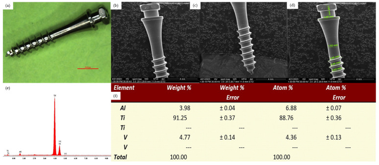
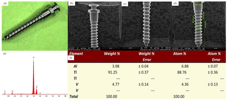
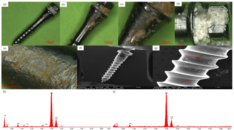
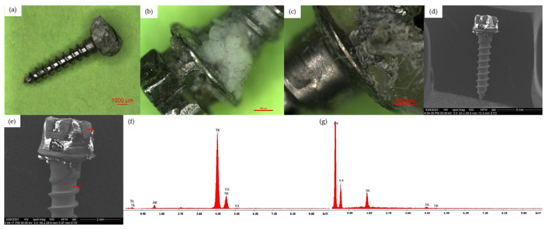
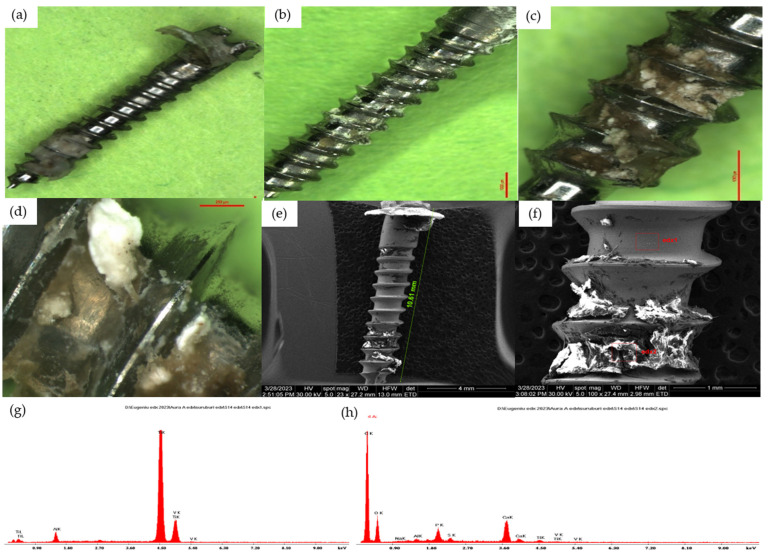
Temporary anchorage devices (TADs) are orthodontic mini-implants with remarkable characteristics that, once inserted, present mechanical retention (primary stability) without the process of bone osseointegration. However, interaction with the biological environment may cause changes in the morphology of the external surface of dental TADs. In this study, we used 17 TADs made of aluminum-vanadium titanium alloy, produced by two companies, which were analyzed through optical microscopy after being removed from the patients during orthodontic treatment. We evaluated the changes that appeared on the TADs' surfaces after their use in the biological environment, depending on the morphological area in which they were inserted. In our study, we found changes in the morphology of the implant surface, and especially deposits of biological material in all study groups. On all samples examined after clinical use, regardless of the period of use, corrosion surfaces in different locations were observed. Our obtained results support the idea that the biological environment is aggressive for mini-implant structures, always producing changes to their surface during their clinical use.

Keywords: bacterial plaque; biocompatibility; corrosion analysis; orthodontic mini-implants; surface morphology changes; temporary anchorage devices.


Conflict of interest statement

The authors declare no conflicts of interest.

Figures

Figure 1
Figure 1
Surface analysis of an unused Dual Top Anchor System implant. (a) Overview aspect seen by optical microscopy, 10×. (b) SEM aspect of the cervical half, Z1-Z2 areas. (c) SEM aspect of the apical half, Z2-Z4 areas. (d) SEM aspect of the cervical half, Z1-Z2 areas with measurements. (e) EDX analysis of the implant surface. (f) Percentage elemental analysis from the implant surface.
Figure 2
Figure 2
Surface analysis of an unused OrthAnchor System implant. (a) Overview aspect seen by optical microscopy, 10×. (b) SEM aspect of the cervical half, Z1-Z3 areas. (c) SEM aspect of the apical half, Z2-Z4 areas. (d) SEM aspect of the implant, Z2-Z4 areas with measurements. (e) EDX analysis of the implant surface. (f) Percentage elemental analysis from the implant surface.
Figure 3
Figure 3
(a) Optical microscopy overview aspect of a Group 1 TAD, 10×. (b) Optical microscopy aspect of a Group 1 TAD, 15×; Z2—cervical surface. (c) Optical microscopy aspect of a Group 1 TAD, 30×; Z2—cervical surface. (d) Optical microscopy aspect of a Group 1 TAD, 45×; Z1—surface of the mini-implant head. (e) Optical microscopy aspect of a Group 1 TAD, 45×; Z2—cervical surface. (f) SEM overview aspect of a Group 1 TAD. (g) SEM aspect of the middle part Z3 area of a Group 1 TAD. (h) EDX analysis from the Z3 area of a Group 1 TAD surface. (i) EDX analysis from the Z4 area of a Group 1 TAD surface.
Figure 4
Figure 4
(a) Optical microscopy overview aspect of a Group 2 TAD, 10×. (b) Optical microscopy aspect of Group 2 TAD, 30×; Z1-Z2—cervical surface. (c) Optical microscopy aspect of Group 2 TAD, 45×; Z2—cervical surface. (d) SEM overview aspect of a Group 2 TAD. (e) SEM aspect of the head half, Z1-Z2 areas of a Group 2 TAD. (f) EDX analysis from the Z1 area of a Group 2 TAD surface. (g) EDX analysis from the Z2 area of a Group 2 TAD surface.
Figure 5
Figure 5
(a) Optical microscopy overview aspect of a Group 3 TAD, 10×. (b) Optical microscopy aspect of Group 3 TAD, 15×; Z2-Z4 areas of a Group 3 TAD. (c) Optical microscopy aspect of group 3 TAD, 30×; Z3-Z4 areas of a Group 3 TAD. (d) Optical microscopy aspect of Group 3 TAD, 75×; Z4 areas of a Group 3 TAD. (e) SEM overview aspect of a Group 3 TAD. (f) SEM analysis from the Z4 area of a Group 3 TAD surface. (g) EDX analysis of a clean zone from the Z4 area of a Group 3 TAD surface. (h) EDX analysis of a covered zone from the Z4 area of a Group 3 TAD surface.

References

    1. Łyczek J., Kawala B., Antoszewska-Smith J. Influence of antibiotic prophylaxis on the stability of orthodontic microimplants: A pilot randomized controlled trial. Am. J. Orthod. Dentofac. Orthop. 2018;153:621–631. doi: 10.1016/j.ajodo.2017.11.025. - DOI - PubMed
    1. Giri M., Sabapathy K., Govindasamy B., Rajamurugan H. Evaluation of insertion torque and surface integrity of zirconia-coated titanium mini screw implants. J. World Fed. Orthod. 2020;9:13–17. doi: 10.1016/j.ejwf.2020.01.002. - DOI - PubMed
    1. Becker K., Pliska A., Busch C., Wilmes B., Wolf M., Drescher D. Efficacy of orthodontic mini implants for en masse retraction in the maxilla: A systematic review and meta-analysis. Int. J. Implant Dent. 2018;4:35. doi: 10.1186/s40729-018-0144-4. - DOI - PMC - PubMed
    1. Moldoveanu A., Nicolescu M.I., Bucur M.V., Moldoveanu G.G., Funieru C., Neagoe I.V., Manda G., Ioana T.R., Ciocan L.T. In vitro study of the orthodontic mini-implants influence on the growth of human osteoblasts. Rom. J. Morphol. Embryol. 2021;62:785–792. doi: 10.47162/RJME.62.3.16. - DOI - PMC - PubMed
    1. Ramírez-Ossa D.M., Escobar-Correa N., Ramírez-Bustamante M.A., Agudelo-Suárez A.A. An Umbrella Review of the Effectiveness of Temporary Anchorage Devices and the Factors That Contribute to Their Success or Failure. J. Evid. Based Dent. Pract. 2020;20:101402. doi: 10.1016/j.jebdp.2020.101402. - DOI - PubMed

LinkOut - more resources