. 2024 Aug 30;15(9):251.
doi: 10.3390/jfb15090251.

The Impact of the Methacrylation Process on the Usefulness of Chitosan as a Biomaterial Component for 3D Printing

Affiliations

The Impact of the Methacrylation Process on the Usefulness of Chitosan as a Biomaterial Component for 3D Printing

Marta Klak et al. J Funct Biomater. .

Abstract

Chitosan is a very promising material for tissue model printing. It is also known that the introduction of chemical modifications to the structure of the material in the form of methacrylate groups makes it very attractive for application in the bioprinting of tissue models. The aim of this work is to study the characteristics of biomaterials containing chitosan (BCH) and its methacrylated equivalent (BCM) in order to identify differences in their usefulness in 3D bioprinting technology. It has been shown that the BCM material containing methacrylic chitosan is three times more viscous than its non-methacrylated BCH counterpart. Additionally, the BCM material is characterized by stability in a larger range of stresses, as well as better printability, resolution, and fiber stability. The BCM material has higher mechanical parameters, both mechanical strength and Young's modulus, than the BCH material. Both materials are ideal for bioprinting, but BCM has unique rheological properties and significant mechanical resistance. In addition, biological tests have shown that the addition of chitosan to biomaterials increases cell proliferation, particularly in 3D-printed models. Moreover, modification in the form of methacrylation encourages reduced toxicity of the biomaterial in 3D constructs. Our investigation demonstrates the suitability of a chitosan-enhanced biomaterial, specifically methacrylate-treated, for application in tissue engineering, and particularly for tissues requiring resistance to high stress, i.e., vascular or cartilage models.

Keywords: biomaterial; bioprinting; chitosan; extracellular matrix; methacrylation; tissue engineering; tissue regeneration.


Conflict of interest statement

Authors Marta Klak, Katarzyna Kosowska, Milena Czajka, Magdalena Dec, Sylwester Domański, Agnieszka Zakrzewska, and Michał Wszoła are employed by the company Polbionica Ltd. The other authors declare no conflicts of interest.

Figures

Figure 1
Figure 1
Diagram showing modifications of chitosan and its applications in medicine.
Figure 2
Figure 2
The scheme of the methacrylation process.
Figure 3
Figure 3
The models of printable structures to evaluate printability in (A) the fiber fusion test and (B) the fiber collapse test.
Figure 4
Figure 4
Stack plot of 1H NMR spectra collected for representative samples of CHIMA.
Figure 5
Figure 5
Results of rheology testing, where (A) is the dependence of the complex modulus on temperature and (B) is the complex modulus for BCH (biomaterial with chitosan) and BCM (biomaterial enriched with methacrylated chitosan).
Figure 6
Figure 6
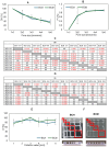
Results of printability testing, where (A) is the diffusion rate, (B) is the printability, and (E) is the collapse area factor for BCH (biomaterial with chitosan) and BCM (biomaterial enriched with methacrylated chitosan). In picture (F), the photos of printed models are shown. Tables (C,D) present the statistical analysis of individual variants of printability experimental results. Statistically significant values are marked in red. In the case of the collapse area factor, the differences were statistically significant for BCM 3 vs. BCH 1 (p = 0.029), BCM 3 vs. BCH 4 (p = 0.0461), BCM 3 vs. BCH 5 (p = 0.0060), BCM 3 vs. BCH 6 (p = 0.0023), BCM 3 vs. BCM 1 (p = 0.0046), and BCM 4 vs. BCM 3 (p = 0.0274). For other data sets, the differences were statistically insignificant.
Figure 7
Figure 7
Results of mechanical testing. (A) The dependence of stress on the strain of test samples: blue color indicates BCH (biomaterial with chitosan) and green color indicates BCM (biomaterial enriched with methacrylated chitosan). (B) Photos of the printed and destroyed BCH and BCM structures. (C) mechanical parameters: the blue color indicates the mechanical strength values and the Young’s modulus values for BCH (biomaterial with chitosan) and the green color indicates the mechanical parameters for BCM (biomaterial enriched with methacrylated chitosan).
Figure 8
Figure 8
Results of water absorption, swelling test, and the degree of degradation for the tested materials. The average mass of mg of water per mg of sample in the (A) water absorption and (B) swelling test for BCH (biomaterial with chitosan) and BCM (biomaterial enriched with methacrylated chitosan); enzymatic degradation (D) after 24 h, 7 days, 14 days, and 21 days; and non-enzymatic degradation result (E) after the same. The tables (C,FH) present the statistical analysis of individual variants of the water absorption experimental results and degradation after time. Statistically significant values are marked in red. In the case of the swelling test, the differences were statistically insignificant.
Figure 9
Figure 9
Microscopic imaging of RFP-HDFCs-Neo cells cultured on biomaterials and as a test model of printed constructs. Imaging was performed 24 h and 7 days after preparation of the constructs.. The images were made with an Olympus IX83 microscope (Olympus, PA, USA) in the bright field (BF) and with the use of red fluorescent lights (TRITC-tetramethylrhodamine). Imaging was performed at both 10× (cell-covered biomaterials) and 4× (3D-printed models) objective magnifications. Control: cells cultured under standard conditions (37 °C and 5% CO2); BCH: cells cultured on biomaterial with added chitosan; and BCM: cells cultured on biomaterial with added methacrylated chitosan.
Figure 10
Figure 10
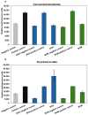
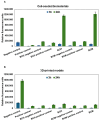
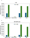
Cytotoxicity of biomaterials. LDH assay was performed as a result of the contact of cells with biomaterials ((A) cell-seeded biomaterials) and after the bioprinting process ((B) 3D-printed models). Negative control: cells cultured under standard conditions; positive control: cells cultured under standard conditions exposed to 0.1% Triton X-100; BCH: cells+ chitosan-biomaterial; BCM: cells+ methacrylated chitosan-biomaterial; BCH/BCM negative control: biomaterials without cells; and BCM/BCH positive control: biomaterials+ cells exposed to 0.1% Triton X-100. Relative luminescence units (RLUs).
Figure 11
Figure 11
Cell proliferation as a result of exposure to biomaterials. (A) Cells cultured on the surface of biomaterials. (B) 3D-printed models. Negative control: cells cultured under standard conditions; positive control: cells cultured under standard conditions exposed to 0.1% Triton X-100; BCH: cells+ chitosan-biomaterial; BCM: cells+ methacrylated chitosan-biomaterial; BCH/BCM negative control: biomaterials without cells; and BCM/BCH positive control: biomaterials+ cells exposed to 0.1% Triton X-100. Relative fluorescence units (RFUs).
Figure 12
Figure 12
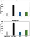
Cytotoxicity of biomaterial extract. LDH assay was performed with the use of L-929 (in accordance with standard PN-EN ISO 10993-5:2009) (A) and RFP-HDFCs-Neo (B) cells. Negative control: cells cultured under standard conditions (37 °C, 5% CO2); positive control: cells cultured under standard conditions (37 °C, 5% CO2) exposed to 0.1% Triton X-100; BCH: cells+ chitosan-biomaterial extracts; and BCM: cells+ methacrylated chitosan-biomaterial extracts.
Figure 13
Figure 13
Cell proliferation after exposure to biomaterial extract. Alamar Blue assay was performed with the use of L-929 (in accordance with standard PN-EN ISO 10993-5:2009) (A) and RFP-HDFCs-Neo (B) cells. Negative control: cells cultured under standard conditions (37 °C, 5% CO2); positive control: cells cultured under standard conditions (37 °C, 5% CO2) exposed to 0.1% Triton X-100; BCH: cells+ chitosan-biomaterial extracts; and BCM: cells+ methacrylated chitosan-biomaterial extracts.
Figure 14
Figure 14
Expression of selected genes in RFP-HDFCs-Neo cells after exposure to biomaterials compared to cells cultured under standard conditions (defined as 1-fold change). (A) Gene expression of cells cultured on the surface of biomaterials; (B) gene expression of cells in 3D-printed models. BCH: cells+ chitosan-based biomaterial; BCM: cells+ methacrylated chitosan-based biomaterial.

Similar articles

References

    1. Kołodziejska M., Jankowska K., Klak M., Wszoła M. Chitosan as an Underrated Polymer in Modern Tissue Engineering. Nanomaterials. 2021;11:3019. doi: 10.3390/nano11113019. - DOI - PMC - PubMed
    1. Wang J., Cui Z., Maniruzzaman M. Bioprinting: A Focus on Improving Bioink Printability and Cell Performance Based on Different Process Parameters. Int. J. Pharm. 2023;640:123020. doi: 10.1016/j.ijpharm.2023.123020. - DOI - PubMed
    1. Gungor-Ozkerim P.S., Inci I., Zhang Y.S., Khademhosseini A., Dokmeci M.R. Bioinks for 3D Bioprinting: An Overview. Biomater. Sci. 2018;6:915–946. doi: 10.1039/C7BM00765E. - DOI - PMC - PubMed
    1. Murphy S.V., Atala A. 3D Bioprinting of Tissues and Organs. Nat. Biotechnol. 2014;32:773–785. doi: 10.1038/nbt.2958. - DOI - PubMed
    1. Sun W., Starly B., Daly A.C., Burdick J.A., Groll J., Skeldon G., Shu W., Sakai Y., Shinohara M., Nishikawa M., et al. The Bioprinting Roadmap. Biofabrication. 2020;12:022002. doi: 10.1088/1758-5090/ab5158. - DOI - PubMed

LinkOut - more resources