. 2024 Aug 24;22(9):382.
doi: 10.3390/md22090382.

Mutagenesis of the Peptide Inhibitor of ASIC3 Channel Introduces Binding to Thumb Domain of ASIC1a but Reduces Analgesic Activity

Affiliations

Mutagenesis of the Peptide Inhibitor of ASIC3 Channel Introduces Binding to Thumb Domain of ASIC1a but Reduces Analgesic Activity

Timur A Khasanov et al. Mar Drugs. .

Abstract

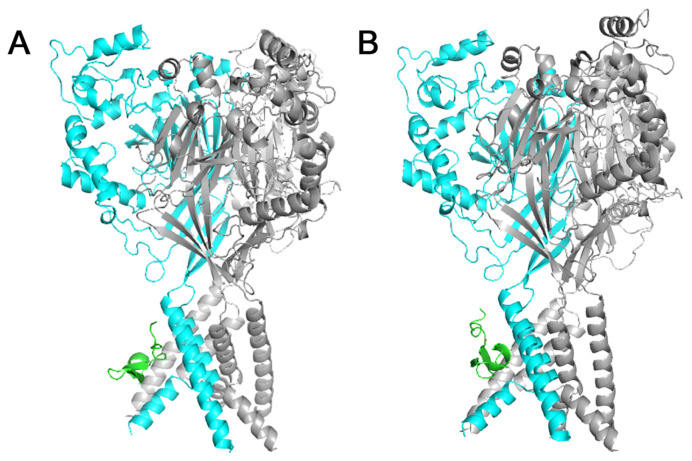
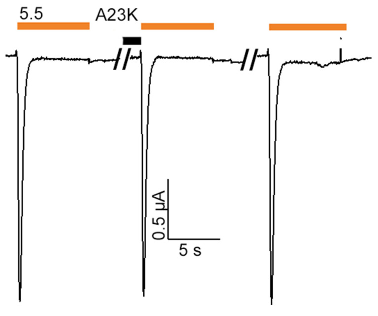
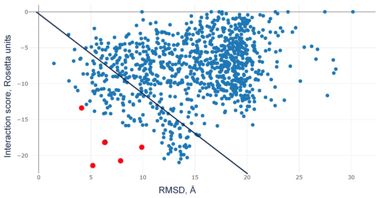
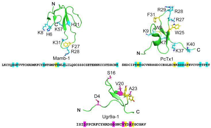
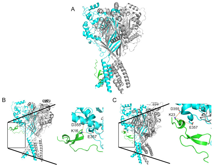
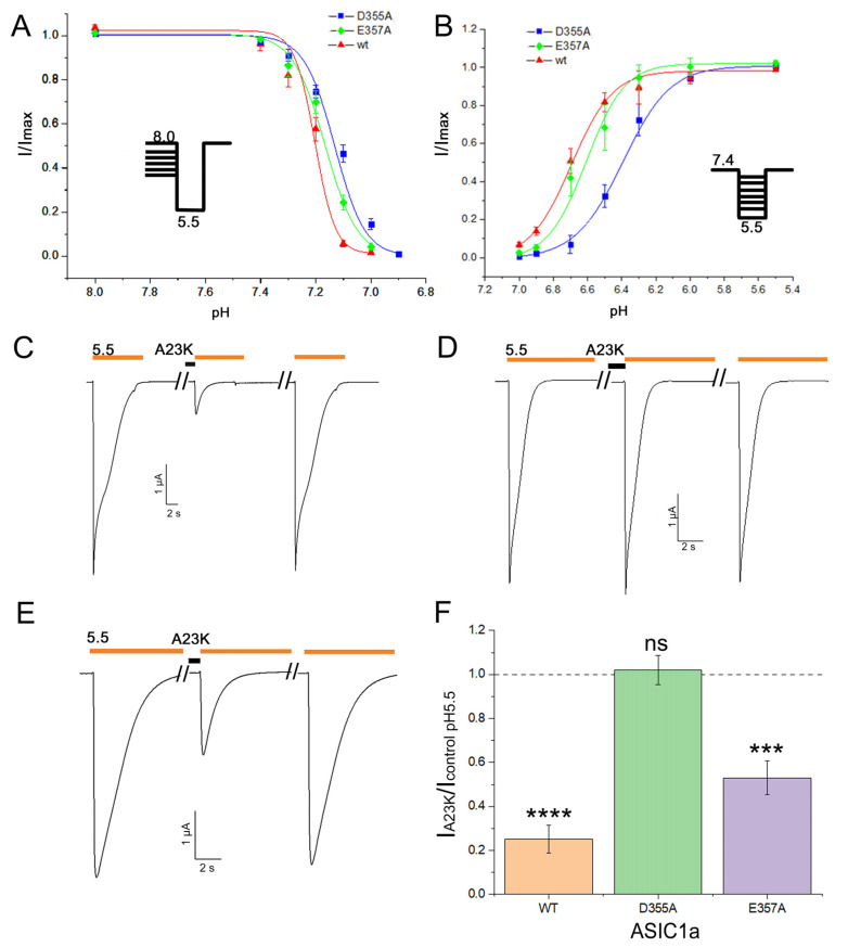
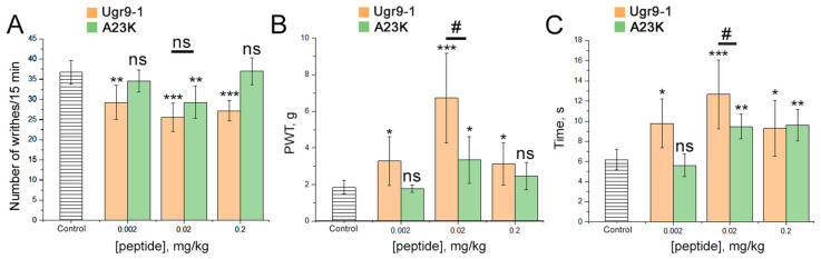
Acid-sensing ion channels (ASICs), which act as proton-gating sodium channels, have garnered attention as pharmacological targets. ASIC1a isoform, notably prevalent in the central nervous system, plays an important role in synaptic plasticity, anxiety, neurodegeneration, etc. In the peripheral nervous system, ASIC1a shares prominence with ASIC3, the latter well established for its involvement in pain signaling, mechanical sensitivity, and inflammatory hyperalgesia. However, the precise contributions of ASIC1a in peripheral functions necessitate thorough investigation. To dissect the specific roles of ASICs, peptide ligands capable of modulating these channels serve as indispensable tools. Employing molecular modeling, we designed the peptide targeting ASIC1a channel from the sea anemone peptide Ugr9-1, originally targeting ASIC3. This peptide (A23K) retained an inhibitory effect on ASIC3 (IC50 9.39 µM) and exhibited an additional inhibitory effect on ASIC1a (IC50 6.72 µM) in electrophysiological experiments. A crucial interaction between the Lys23 residue of the A23K peptide and the Asp355 residue in the thumb domain of the ASIC1a channel predicted by molecular modeling was confirmed by site-directed mutagenesis of the channel. However, A23K peptide revealed a significant decrease in or loss of analgesic properties when compared to the wild-type Ugr9-1. In summary, using A23K, we show that negative modulation of the ASIC1a channel in the peripheral nervous system can compromise the efficacy of an analgesic drug. These results provide a compelling illustration of the complex balance required when developing peripheral pain treatments targeting ASICs.

Keywords: acid-sensing ion channel; analgesic effect; molecular docking; mutagenesis; pain models; peptide ligand.


Conflict of interest statement

The authors declare no conflicts of interest.

Figures

Figure A1
Figure A1
Molecular docking of Ugr9-1 mutants D4K (A) and V20K (B) to rat ASIC1a channel in close state. The representation showcases the three channel subunits (cyan and gray levels) and peptides (green) depicted in ribbon form. The D4K and V20K mutants are observed positioned within the region of transmembrane domains.
Figure A2
Figure A2
Reverse phase HPLC purification steps of recombinant Ugr9-1 (A,B) and A23K (C,D) peptides utilizing a linear gradient of acetonitrile (represented by the blue straight line). The fraction containing the target product is highlighted within a gray rectangle. Absorbance at 210 nm is depicted by a black curve, absorbance at 280 nm is represented by a blue curve.
Figure A3
Figure A3
Lack of effect of mutant A23K peptide on rat ASIC1b channel activity. Representative current traces of homomeric ASIC1b channels activated at pH 5.5 (indicated by orange bars). Traces depict the channel activity before peptide application, after 30 s of preapplication of A23K peptide at a concentration of 50 µM (black bar), and after washout, respectively.
Figure A4
Figure A4
Scatter plot of interaction score (Rosetta units) vs. RMSD (Å) of complex A23K-rASIC1a. Red dots represent 5 models with highly negative ratio of interaction score to RMSD among all generated models (blue dots). Black line represents values where the ratio equals to −1.
Figure 1
Figure 1
The structures of three peptides: Mamb-1 sourced from snake venom, PcTx1 from tarantula venom, and Ugr9-1 from sea anemone venom. The primary and spatial structures of the peptides inhibiting ASIC channels: Mamb-1, PcTx1, and Ugr9a-1 (PDB codes 1MJU, 2KNI, and 2LZO, respectively). Positively charged residues important for inhibition of the ASIC1a channel are highlighted in blue in Mam-1 and PcTx1. Residues earmarked for substitution in the Ugr9-1 structure are highlighted in pink. Additionally, tyrosine residues, putatively forming a basic-aromatic cluster within the Ugr9-1 structure, are denoted in yellow.
Figure 2
Figure 2
Molecular docking of the wild-type Ugr9-1 (A) alongside its mutants S16K (B) and A23K (C) to the rat ASIC1a channel in closed state. The representation showcases the three channel subunits (cyan and gray levels) and peptides (green) depicted in ribbon form. In (A), the Ugr9-1 is observed positioned within the region of transmembrane domains. Conversely, in mutants S16K (B) and A23K (C), lysine residues at positions 16 and 23, respectively, are presumed to instigate interactions with residues D355 and E357 of the channel. Distances are delineated by orange dotted lines.
Figure 3
Figure 3
The effect of mutant peptides S16K and A23K on rat ASIC1a and ASIC3 channels. (A) Representative current traces demonstrating the effects of 12.5 µM S16K and A23K on ASIC1a as well as the lack of effect observed with 50 µM Ugr9-1 on ASIC1a. (B) Dose–response curves for the inhibitory effect of mutant peptides on ASIC1a. Each data point represents data from 5 cells. (C) Representative current traces of Ugr9-1, S16K, and A23K at a concentration of 12.5 µM on ASIC3. (D) Dose–response curves revealing the inhibitory effect of both Ugr9-1 and mutant peptides on ASIC3. Each point represents data from 5 cells. The depicted currents were induced by a pH 5.5 stimulus following a conditioning pH of 7.4, with peptides applied for 30 s before the pH stimulus. The holding potential (Vh) was −50 mV, and the time interval between applications ranged from 1 to 2 min. Data are presented as mean ± S.E.M.
Figure 4
Figure 4
The effect of the mutant peptide A23K on rat heteromeric ASIC1a/3 channels. (A) Representative current traces demonstrating the effect of 12.5 µM A23K. The depicted currents were induced by a pH 5.5 stimulus following a conditioning pH of 7.4, with the peptide applied for 30 s before the pH stimulus. (B) Dose–response curve for the inhibitory effect of the mutant peptide. Each data point represents data from 6 cells. Data are presented as mean ± S.E.M.
Figure 5
Figure 5
The pivotal role of residue D355 in the ASIC1a channel for the action of the A23K peptide ligand. (A,B) pH dependence of steady-state desensitization (A) and activation (B) in mutant channels (D355A and E357A) compared to the wild-type channel (wt). Each data point represents results from 5 cells. (CE) Representative current traces illustrating the effects of A23K at a concentration of 10 µM on wt (C), D355A (D), and E357A (E) channels. Currents were evoked by a pH 5.5 stimulus following a conditioning pH of 7.4, with the peptide applied for 30 s preceding the pH stimulus. (F) Bar graph quantifying the effects depicted in (C,D) as a percentage of the corresponding control currents (n = 5). A value of 1.0, represented by a dotted line, corresponds to control measurements at pH 5.5 for each form of ASIC1a. The data are expressed as mean ± SEM; *** p < 0.001, **** p < 0.0001, ns non-significant vs. control, unpaired t-test.
Figure 6
Figure 6
Effects of Ugr9-1 and A23K peptides in pain models. Peptides (Ugr9-1–orange bars, A23K–green bars) were administered intramuscularly one hour before testing. (A) The efficacy of peptides in the acetic acid-induced writhing test, evaluated based on the number of writhes following intraperitoneal administration of acetic acid. (B,C) Peptide effectiveness in hyperalgesia induced by complete Freund’s adjuvant assessed by the paw withdrawal threshold (PWT) using von Frey filaments of varying forces (B) and the duration of the inflamed hind paw contact with the hot plate (C). Results are presented as mean ± SD (n = 7 for each group). Statistical significance was determined using one-way ANOVA with post hoc Tukey’s test, denoted as * p < 0.05, ** p < 0.01, *** p < 0.001 for significant differences compared to control; # p < 0.05 for comparisons between experimental groups; and ns—not significant.

Similar articles

Cited by

References

    1. Kellenberger S., Schild L. Epithelial Sodium Channel/Degenerin Family of Ion Channels: A Variety of Functions for a Shared Structure. Physiol. Rev. 2002;82:735–767. doi: 10.1152/physrev.00007.2002. - DOI - PubMed
    1. Kellenberger S., Schild L. International Union of Basic and Clinical Pharmacology. XCI. Structure, Function, and Pharmacology of Acid-Sensing Ion Channels and the Epithelial Na+ Channel. Pharmacol. Rev. 2015;67:1–35. doi: 10.1124/pr.114.009225. - DOI - PubMed
    1. Hung C.-H., Chin Y., Fong Y.-O., Lee C.-H., Han D.-S., Lin J.-H., Sun W.-H., Chen C.-C. Acidosis-related pain and its receptors as targets for chronic pain. Pharmacol. Ther. 2023;247:108444. doi: 10.1016/j.pharmthera.2023.108444. - DOI - PubMed
    1. Hsieh W.-S., Kung C.-C., Huang S.-L., Lin S.-C., Sun W.-H. TDAG8, TRPV1, and ASIC3 involved in establishing hyperalgesic priming in experimental rheumatoid arthritis. Sci. Rep. 2017;7:8870. doi: 10.1038/s41598-017-09200-6. - DOI - PMC - PubMed
    1. Wemmie J.A., Chen J., Askwith C.C., Hruska-Hageman A.M., Price M.P., Nolan B.C., Yoder P.G., Lamani E., Hoshi T., Freeman J.H., et al. The Acid-Activated Ion Channel ASIC Contributes to Synaptic Plasticity, Learning, and Memory. Neuron. 2002;34:463–477. doi: 10.1016/S0896-6273(02)00661-X. - DOI - PubMed

LinkOut - more resources