

Staurosporine as a Potential Treatment for Acanthamoeba Keratitis Using Mouse Cornea as an Ex Vivo Model
- PMID: 39330304
- PMCID: PMC11433162
- DOI: 10.3390/md22090423
Staurosporine as a Potential Treatment for Acanthamoeba Keratitis Using Mouse Cornea as an Ex Vivo Model
Abstract
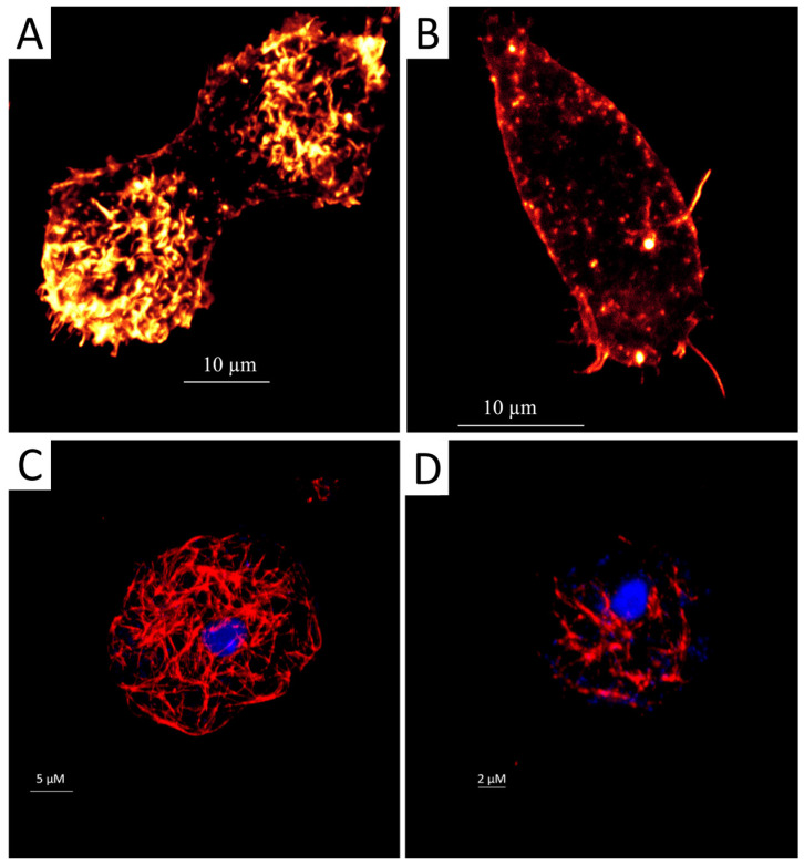
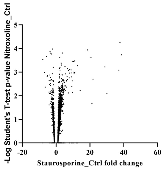
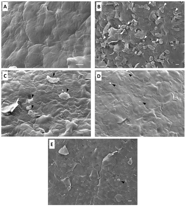
Acanthamoeba is a ubiquitous genus of amoebae that can trigger a severe and progressive ocular disease known as Acanthamoeba Keratitis (AK). Furthermore, current treatment protocols are based on the combination of different compounds that are not fully effective. Therefore, an urgent need to find new compounds to treat Acanthamoeba infections is clear. In the present study, we evaluated staurosporine as a potential treatment for Acanthamoeba keratitis using mouse cornea as an ex vivo model, and a comparative proteomic analysis was conducted to elucidate a mechanism of action. The obtained results indicate that staurosporine altered the conformation of actin and tubulin in treated trophozoites of A. castellanii. In addition, proteomic analysis of treated trophozoites revealed that this molecule induced overexpression and a downregulation of proteins related to key functions for Acanthamoeba infection pathways. Additionally, the ex vivo assay used validated this model for the study of the pathogenesis and therapies of AK. Finally, staurosporine eliminated the entire amoebic population and prevented the adhesion and infection of amoebae to the epithelium of treated mouse corneas.
Keywords: Acanthamoeba; PCD; ex vivo; mouse cornea; proteomic analysis; staurosporine.
Conflict of interest statement
The authors declare no conflicts of interest.
Figures
Similar articles
-
Clinical and histologic evaluations of experimental Acanthamoeba keratitis.Parasitol Res. 2007 Nov;101(6):1621-5. doi: 10.1007/s00436-007-0704-7. Epub 2007 Aug 14. Parasitol Res. 2007. PMID: 17701053
-
Amoebicidal Activity of Poly-Epsilon-Lysine Functionalized Hydrogels.Invest Ophthalmol Vis Sci. 2022 Jan 3;63(1):11. doi: 10.1167/iovs.63.1.11. Invest Ophthalmol Vis Sci. 2022. PMID: 34994769 Free PMC article.
-
Temperature limitation may explain the containment of the trophozoites in the cornea during Acanthamoeba castellanii keratitis.Parasitol Res. 2014 Dec;113(12):4349-53. doi: 10.1007/s00436-014-4109-0. Epub 2014 Sep 10. Parasitol Res. 2014. PMID: 25204727
-
The biology of Acanthamoeba keratitis.Exp Eye Res. 2021 Jan;202:108365. doi: 10.1016/j.exer.2020.108365. Epub 2020 Nov 19. Exp Eye Res. 2021. PMID: 33221372 Free PMC article. Review.
-
The role of the innate and adaptive immune responses in Acanthamoeba keratitis.Arch Immunol Ther Exp (Warsz). 2002;50(1):53-9. Arch Immunol Ther Exp (Warsz). 2002. PMID: 11916309 Review.
References
-
- Henriquez F.L. Review of “Acanthamoeba: Biology and Pathogenesis” by Naveed Ahmed Khan. Parasit. Vectors. 2009;2:16. doi: 10.1186/1756-3305-2-16. - DOI
MeSH terms
Substances
Grants and funding
LinkOut - more resources
Full Text Sources
